Ucell™ 96 Well Cell Culture Plate,Ultra-Low Attachment Surface, U-bottom, Clear, Sterile

$399.00

SKU: L1096UA
Enter the quantity you would like, or drag the panel to find the quantity and price that best for you
Add to QuoteAuthorize OEM & ODM

Product details

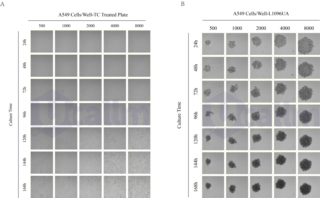
The bottom of the ultra-low adsorption cell culture plate/dish/bottle is attached to the hydrogel with the surface covalent bond, which can minimize the cell attachment, cell activation, protein absorption and enzyme activation; The hydrogel had no cytotoxic effect. It can be used for cell non-adherent culture, organoids, spheroids and other cultures, for the collection and storage of rare samples, and has anti-adherent properties for strongly adhesive cells.

  • Surfaces minimize protein absorption, enzyme activation, and cell activation
  • The surface is non-cytotoxic, bioinert and non-degradable
  • Uniform floor space for easy stacking
  • Irreversible cover with condensing ring for reduced contamination
  • The top of the lid has a stacking ring for easy stacking
  • Irradiation sterilization, SAL10-6
  • No DNase/RNase, no pyrogen, no cytotoxicity

Product characteristics

Label/ Print With plastic label
Closure type Slip-on lid
Base shape U base
Number of wells 96
Growth area 0.32 cm²
Volume of work 0.07-0.2 mL
Vessel type Plate

Specifications

● Size
Width of product85.5 mm
Height of product 14.2 mm
Length of product 127.7 mm
●Material & colours
Product material Polystyrene (PS)
Colour of product Clear
Closure material Polystyrene (PS)
● Purity & certification
Product categoryNo medical device | IVD
Surface Treatment TC-treated
Sterilisation Gamma irradiation
Pyrogen-free Yes
Non-cytotoxicYes
BatchedYes
● Packaging
Qty/Pk1/Pk
Qty/Cs5/Cs

Applications

Formation of Tumor Spheroids of A549 (Human Non-Small Cell Lung Cancer Cells)

Origin and Characteristics of the A549 Cell Line

The A549 cell line was initially isolated in 1972 from the lung tissue of a 58-year-old Caucasian male patient with lung adenocarcinoma[1]. These cells are cancerous derivatives of human alveolar basal epithelial cells and possess unique characteristics that make them a common model for studying lung cancer and other related diseases[2]. A549 cells are adherent and can form monolayers in culture dishes; however, under specific culture conditions, they can also form three-dimensional spheroids[3].  

Applications of the A549 Cell Line

  • Cancer Biology Research
    • The A549 cell line is widely used to study the mechanisms of lung cancer initiation, progression, and metastasis. Researchers utilize this cell line to investigate the roles of specific genes or signaling pathways in lung cancer cells, such as the EGFR, KRAS, and PI3K/AKT pathways[1,2]
  • Drug Screening and Evaluation
    • The A549 cell line is a frequently used model for drug screening and evaluation. Researchers employ it to assess the efficacy of novel anti-cancer drugs and to study drug mechanisms of action and resistance[2].
  • Toxicological Studies
    • The A549 cell line can also be used to evaluate the toxicity of environmental pollutants or chemicals on lung cells[1]. By measuring indicators such as cell viability, apoptosis, and inflammatory response after exposure to various substances, the potential hazards of these compounds can be assessed.
  • Three-Dimensional (3D) Cell Culture
    • The A549 cell line is suitable for 3D cell culture and can form tumor spheroids. Compared to traditional two-dimensional (2D) culture, 3D culture better simulates the in vivo tumor microenvironment, providing a more realistic model for studying tumor growth, invasion, and metastasis[3].
  • Other Applications
    • Beyond the applications mentioned above, the A549 cell line is also used in research on viral infections, gene therapy, and the biological effects of nanomaterials[1].
 

Morphological Observation and Live Cell Station Detection of A549 at Different Densities -Ultra-Low Attachment Plate (U-bottom)

A549 cells cultured on Ultra-Low Attachment Surface forms tumor spheroids. A549 were seeded onto the T-25TCT (A) and Ultra-Low Attachment Surface (B)vessels.The cells were cultured for 24, 48,72, 96, 120, 144 and 168 hours and a representative image was taken using an Ucallm living image station. Scale barsrepresent 100 μm..

Figure 1. MCF-7 cells cultured on CorningUltra-Low Attachment Surface forms tumorspheroids. MCF-7 were seeded onto the T-25TCT (A) and Ultra-Low Attachment Surface (B)vessels.The cells were cultured for 72 hoursand a representative image was taken usingan AMG EVOS® Fl microscope. Scale barsrepresent 200 um.

Figure 1:Live imaging of A549 spheroids in Ucallm® Ultra low attachment cell culture plate(#L1096UA) compared to two benchmark products on days 1, 3, and 4. Scale bar: 100 μm.

Method

Cell Culture and Treatment
Culture Conditions: 89% DMEM/F12 medium, 10% FBS and 1% P/S
Cell Thawing
  1. Retrieve A549 cells from liquid nitrogen and quickly place them in a 37℃ water bath, gently shaking the cryovial to thaw the freezing medium.
  2. After thawing, transfer the cells to a centrifuge tube containing 3 mL of medium, centrifuge to pellet the cells (1000 rpm, 5 min, room temperature), and discard the supernatant.
  3. Resuspend the cells in complete medium, seed them into a culture dish, gently pipet to mix, and culture in a normal incubator at 37℃, 21% O2, 5% CO2.
Cell Passaging
  1. When cell density reaches 80%, passage the cells.
  2. Discard the medium and wash once with PBS.
  3. Add 1-2 mL of 0.25% trypsin to digest the cells at 37℃for 2-5 min.Observe under a microscope; digestion is complete when cells separate and become rounded.
  4. Quickly discard the trypsin, add complete medium, pipet to create a single-cell suspension, and passage at a ratio of 1:2 to 1:4. Expand the culture in a normal incubator at 37℃, 21% O2, 5% CO2.
Cell Seeding
Take A549 cells in the logarithmic growth phase with good growth status. Seed them into a 96-well U-bottom ultra-low attachment plate at densities of 500, 1000, 2000, 4000, and 8000 cells/well (one plate total), with 5 replicate wells per density. Add 100 μL of sterile PBS to the peripheral wells of the plate. Place the 96-well U-bottom ultra-low attachment plate in the live cell station for culture and imaging. Note: The live cell station was installed in the CO2 incubator (CTCC, PH-C-30L) and prewarmed for 30 min, maintaining conditions at 37℃, 21% O2, 5% CO2, and saturated humidity.
Medium Change
For the first 3 days, the initial number of A549 cells is low and metabolism is relatively slow; change the medium every 24-48 hours. From days 4-7, as cell proliferation increases and density gradually rises, metabolism accelerates, requiring medium changes every 1-2 days (Note: Medium changes are also performed synchronously for the plate in the live cell station. The medium change frequency for the 500, 1000, and 2000 cells/well densities is lower than that for the 4000 and 8000 cells/well densities). During medium change, carefully transfer the cell spheroids gently using a 200 μL yellow pipette tip to a 35 mm cell culture dish containing 1 mL of complete medium. Transfer the spheroids from all 6 replicate wells of one density at once. Then, aspirate and discard the old medium from the wells, rinse the wells 2-3 times with 100 μL/well of PBS. Subsequently, use the 200 μL yellow pipette tip to transfer the cell spheroids back into the wells one by one, maintaining a medium volume of 100 μL per well.
Cell Imaging
  • Live Cell Station Imaging
Place the cell plate into the sample holder of the live cell monitoring system. Set the imaging parameters:
  1. Objective: 10X;
  2. Channel selection: Brightfield (BF), light intensity 2%, adjust exposure (within 1000 µs) and gain to increase brightness;
  3. Confirm cell status: Using the navigation interface, check the focal plane for each well, starting with the focal plane of well C3;
  4. Scan type: 1 field of view per well;
  5. Scan interval: Scan every 1 hour, continuously for 168 hours, until the experiment is completed.
  • Optical Microscope Imaging
Every 24 hours, take the cell plate out of the incubator and place it under the optical microscope. Set the imaging parameters:
  1. Objective: 20X, 40X;
  2. Channel selection: Brightfield (BF), Phase Contrast: BF, light intensity 20%, adjust exposure (within 1000 µs) and gain to increase brightness;
  3. Confirm cell status: By moving the cell plate, adjust the field of view for the cells in each well to the center;
  4. Imaging: Capture one 200X image and one 400X image per well.
 

Materials and Instruments

Table 1 Main equipment
Name Manufacturer Catalog Number
Micropipette ThermoFisher
Clean Bench Suzhou Group Antai Air Technology Co., Ltd. SW-CJ-1FD
CO2 Incubator CTCC PH-C-30L
Inverted Microscope OLYMPUS IX73
Optical Microscope Shanghai Precision Instruments XDS-1A
Low-Speed Centrifuge Shanghai Luxiangyi Instruments TDZ4B-WS
96-well Ultra-Low Attachment(U-bottom)Cell Culture Plate
Table 2 Main reagents
Name Manufacturer Catalog Number
A549 Cells CTCC HCL-0023
DMEM/F12 Hyclone SH30023.01B
FBS Lonsera A511-001
Penicillin-Streptomycin Solution Biosharp BL505A
0.25% Trypsin CTCC 002PI
PBS Buffer Solution CTCC F002
 

References

  1. Eşiyok N, Heide M. The SVZ stem cell niche–components, functions, and in vitro modelling [J]. Frontiers in Cell and Developmental Biology, 2023, 11.
  2. Papapostolou I, Bochen F, Peinelt C, et al. A Simple and Fast Method for the Formation and Downstream Processing of Cancer-Cell-Derived 3D Spheroids: An Example Using Nicotine-Treated A549 Lung Cancer 3D Spheres [J]. Methods and Protocols, 2023, 6(5): 94.
  3. [Oszvald Á, Szvicsek Z, Pápai M, et al. Fibroblast-Derived Extracellular Vesicles Induce Colorectal Cancer Progression by Transmitting Amphiregulin [J]. Frontiers in Cell and Developmental Biology, 2020, 8.
Please login to post questions

Back to Top
Product has been added to your cart

Dr. Michael J. Williams

Hello! Welcome to the Ucallm. Click the button below to send me an email.Let me know your requirements and I’ll provide you with a preferential quote.

Powered by WpChatPlugins