
When it comes to cell culture experiments, achieving the right cell density is crucial for successful outcomes. For researchers using 8 well chamber slides, understanding how many cells to plate is essential for accurate microscopic analysis.
We recognize that different cell types and experimental applications require specific cell densities. The 8 well chamber slide, with its surface area of approximately 0.7 cm² per well, offers a versatile tool for various laboratory experiments.
To ensure consistent and reproducible results, it’s vital to follow precise cell plating techniques. In this article, we will guide you through the technical specifications of chamber slides and provide recommendations for achieving optimal cell density.
Key Takeaways
- Understand the optimal cell plating density for different cell types.
- Learn the technical specifications of 8 well chamber slides.
- Discover how to achieve consistent and reproducible results.
- Explore the importance of proper cell plating techniques.
- Find guidance on experimental applications using chamber slides.
What Are 8 Well Chamber Slides
The 8 well chamber slide is a specialized laboratory consumable designed to facilitate efficient cell culture and analysis. We will explore its structure and components in detail.
Structure and Components of Chamber Slides
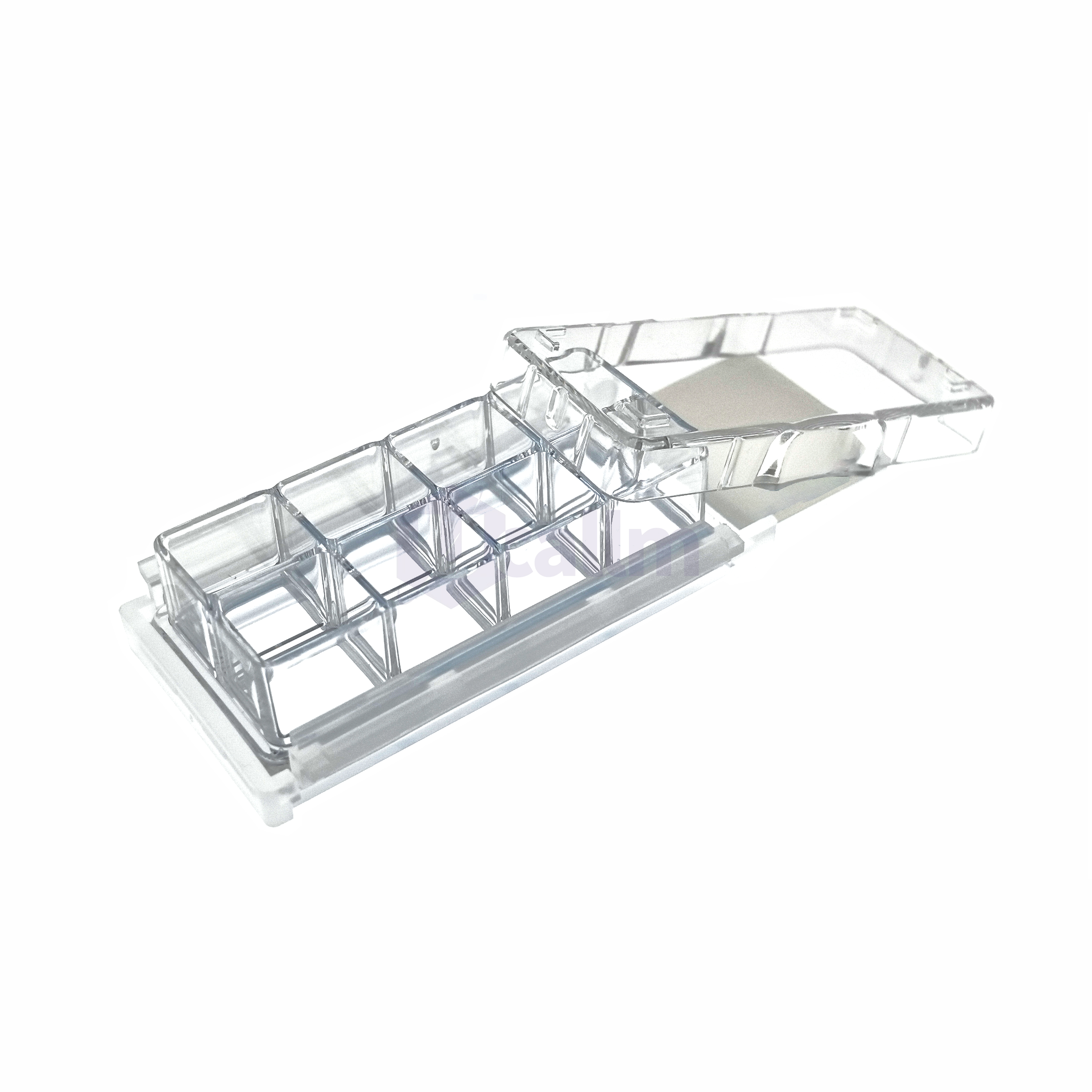
8 well chamber slides are designed to offer the unique benefit of culturing up to 8 different conditions on one standard glass slide. The chamber apparatus uses no adhesive and does not require a special tool for removal. These slides are versatile tools that combine the benefits of cell culture vessels with the convenience of microscope slides for direct analysis without cell transfer.
The design features eight separate wells on a single glass substrate, allowing researchers to conduct multiple experimental conditions simultaneously. Each well functions as an independent culture environment, making these slides ideal for comparative studies and experimental efficiency.
| Feature | Description | Benefit |
|---|---|---|
| 8 Separate Wells | Allows for multiple experimental conditions | Enhances comparative studies and experimental efficiency |
| Glass Substrate | Compatible with standard microscopy | Facilitates direct analysis without cell transfer |
| No Adhesive Used | Simplifies chamber removal | Reduces risk of contamination or damage |
Understanding the details of these slides is essential for proper handling and optimal experimental design in cell biology research.
Surface Area of 8 Well Chamber Slides
The surface area of each well in an 8 well chamber slide is a critical factor in determining cell plating density. When using these slides, it’s essential to understand the available surface area for cell growth.
Significance in Cell Culture Experiments
The surface area of each well is approximately 0.7 cm², a specification that directly influences cell plating decisions. We must consider this limited growth surface when calculating cell density to avoid overcrowding or sparse distribution, both of which can affect experimental outcomes.
- The surface-to-volume ratio in these chamber slides impacts nutrient availability, gas exchange, and the cellular microenvironment.
- Researchers need to balance the relationship between surface area and media volume to ensure optimal cell growth and function.
- Understanding the exact dimensions of these products helps standardize protocols across different experimental setups and laboratory environments.
To illustrate the importance of surface area, let’s consider the following comparison:
| Well Type | Surface Area (cm²) | Recommended Cell Density |
|---|---|---|
| 8 Well Chamber Slide | 0.7 | 1-5 x 10^4 cells |
| 6 Well Plate | 9.6 | 1-5 x 10^5 cells |
As shown, the surface area of the 8 well chamber slide is significantly smaller than that of a 6 well plate, requiring adjustments in cell plating density.
By understanding the surface area of the chamber slide and its implications for cell culture, you can optimize your experimental design and achieve more reliable results with your product.
Recommended Cell Densities by Cell Type

To ensure optimal growth and experimental outcomes, it’s essential to understand the recommended cell densities for different cell types in 8 well chamber slides. Different cell types require specific plating densities to achieve optimal growth and experimental results.
Adherent vs. Suspension Cell Requirements
Adherent cells, such as epithelial cells and fibroblasts, have different requirements compared to suspension cells like immune cells. Epithelial cells typically require 2-5 × 10⁴ cells per well (approximately 3-7 × 10⁴ cells/cm²) to form a confluent monolayer within 24-48 hours.
Fibroblasts, which are larger and more spread out, perform best at 1-3 × 10⁴ cells per well to prevent overcrowding and contact inhibition. In contrast, neuronal cells need lower densities of 0.5-2 × 10⁴ cells per well to allow for proper neurite extension and network formation.
Suspension cells, such as immune cells, may require higher densities of 5-10 × 10⁴ cells per well depending on the specific application and observation timeframe. It’s also important to note that the glass slide base of chamber slides may influence cell attachment differently than standard tissue culture plastic, necessitating optimization for each cell line.
By understanding these requirements, you can optimize your cell culture experiments in 8 well chamber slides. Whether you’re working with adherent or suspension cells, using the right cell density is crucial for achieving reliable and meaningful results.
Calculating the Optimal Cell Number
The process of calculating the optimal cell number involves several key factors. When using 8 well chamber slides, researchers must consider the desired cell density, the surface area of the well, and the specific requirements of their cell type.
Mathematical Formulas for Cell Density Determination
To determine the optimal cell number, you can use a simple formula: desired cell density (cells/cm²) × surface area (0.7 cm²) = cells per well. For most applications, starting with 2-5 × 10⁴ cells per well is recommended.
The calculation should account for the cell doubling time if the experiment spans multiple days. “Slower-growing cells require higher initial seeding densities,” as noted in cell culture guidelines. The glass surface of the product may need pre-coating with extracellular matrix proteins for optimal cell attachment, affecting the effective plating density.
- Consider the final confluence needed for your specific application when adjusting initial cell numbers.
- Pilot experiments with varying cell densities can help determine ideal conditions for each cell type and experimental endpoint.
By carefully calculating the optimal cell number and considering the specific requirements of your experiment, you can ensure reliable and reproducible results.
Standard Cell Concentrations for Different Experiments
The success of cell culture experiments heavily relies on optimizing cell density for specific applications in 8 well chamber slides. Different experimental setups have unique requirements to achieve reliable and reproducible results.
Optimizing Cell Density for Specific Applications
Various cell-based assays and experiments necessitate distinct cell concentrations to meet their specific objectives. For instance:
- Immunofluorescence studies typically require 30-50% confluence (approximately 1-2 × 10⁴ cells per well) to allow clear visualization of individual cellular structures.
- Transfection experiments benefit from higher cell densities of 3-5 × 10⁴ cells per well (70-80% confluence) to maximize transfection efficiency while maintaining cell health.
- Live cell imaging applications often require lower densities of 0.5-1 × 10⁴ cells per well to facilitate tracking of individual cells over time without overcrowding.
- Drug treatment studies may need varying densities depending on the mechanism being studied, with cytotoxicity assays typically using 2-4 × 10⁴ cells per well.
- Co-culture experiments require careful balancing of multiple cell types, with primary cells often plated at 1-3 × 10⁴ cells per well and supporting cells at adjusted ratios.
The detailed description of cell culture conditions, including precise cell numbers, is essential for experimental reproducibility and valid scientific conclusions. By optimizing cell density, researchers can ensure that their experiments are conducted under optimal conditions, leading to more reliable and interpretable results.
In conclusion, understanding the standard cell concentrations for different experiments is crucial for achieving success in cell culture studies. By adhering to these guidelines, researchers can optimize their experimental setups to obtain high-quality data.
Preparing Cell Suspensions for Accurate Plating
To achieve reliable results, it’s essential to prepare cell suspensions correctly for plating in chamber slides. This process involves several critical steps that ensure accurate and consistent cell distribution across the wells.
First, accurate cell counting is necessary to determine the concentration of the stock cell suspension. You can use a hemocytometer or an automated cell counter for this purpose. Additionally, assessing cell viability using methods like trypan blue exclusion or fluorescent viability dyes is crucial to ensure that only live cells are counted.
Cell Counting and Viability Assessment
When preparing a homogeneous cell suspension, gentle but thorough mixing is required to prevent cell clumping. This is vital because clumping can lead to uneven distribution across the chamber slide wells. You must also ensure that the volume of cell suspension added to each well is consistent, typically between 200-400 μL, to maintain uniform growth conditions across all experimental groups.
Handling these specialized products requires care during the plating process to avoid cross-contamination between wells and to ensure the chamber remains securely attached to the slide base.
By following these guidelines, you can ensure that your cell suspensions are prepared accurately for plating in chamber slides, leading to more reliable and consistent experimental results.
Key Features of 8 Well Chamber Slides

8 well chamber slides offer a range of benefits, including improved microscopic observation and reduced cross-contamination. These laboratory tools are designed to provide researchers with a versatile and efficient means of conducting cell-based assays.
Advantages for Cell-Based Assays
The design of 8 well chamber slides includes several key features that enhance their utility in cell culture experiments. These features include:
- Removable Chambers: Allow for direct microscopic examination of cultured cells without transfer, reducing cell loss and maintaining spatial relationships.
- Optical Quality Glass Base: Provides superior imaging capabilities compared to standard plastic culture vessels, particularly for high-resolution and fluorescence microscopy.
- Alphanumeric Labeling: Facilitates sample tracking and experimental organization by labeling each well.
- Cross-Contamination Prevention: The innovative design prevents cross-contamination between wells, making these slides ideal for comparing multiple treatment conditions simultaneously.
- Leak-Proof Sealing Technology: Maintains sterility while allowing for easy chamber removal without damaging the cultured cells.
- Standardized Dimensions: Ensures compatibility with most microscope stages and imaging systems for seamless workflow integration.
By incorporating these features, 8 well chamber slides provide a reliable and efficient solution for researchers conducting cell-based assays. The combination of a glass base, removable chambers, and alphanumeric labeling makes these slides an essential tool in various laboratory applications.
Cell Distribution Techniques for Even Coverage
Even cell distribution across the chamber slide is essential for obtaining reliable and reproducible results in cell culture studies. We will explore various techniques to achieve uniform cell distribution and discuss methods to prevent cell clumping.
Methods to Prevent Cell Clumping
To ensure even cell distribution, it’s crucial to prevent cell clumping. Several techniques can be employed:
- The “figure-eight” motion technique involves gently moving the chamber slide in a figure-eight pattern immediately after adding the cell suspension.
- Allowing the slide to rest flat for 10-15 minutes in the biosafety cabinet before transferring it to the incubator helps cells settle evenly.
- Pre-warming media and the chamber slide to 37°C before cell plating prevents thermal convection currents.
- Coating the surface with appropriate attachment factors promotes uniform adhesion.
- Avoiding air bubbles during plating is essential to prevent cell-free zones.
Let’s delve deeper into these techniques. The “figure-eight” motion helps distribute cells evenly across the chamber slide. Pre-warming the media and slide to 37°C is also crucial as it prevents thermal shock to the cells, thereby promoting even distribution in cell culture applications.
| Technique | Description | Benefits |
|---|---|---|
| Figure-Eight Motion | Gently moving the chamber slide in a figure-eight pattern | Even cell distribution |
| Pre-warming | Pre-warming media and chamber slide to 37°C | Prevents thermal convection currents |
| Surface Coating | Coating the glass slide surface with attachment factors | Promotes uniform cell adhesion |
By implementing these techniques, researchers can achieve a uniform cell distribution across the chamber slide, which is critical for the success of cell culture experiments.
Preventing Common Cell Plating Errors
To achieve reliable results in cell culture experiments using 8 well chamber slides, it’s crucial to avoid common plating mistakes. Several factors can influence the outcome of your cell culture, including cell density, distribution, and handling of the slide assembly.
Troubleshooting Cell Adhesion Issues
Cell adhesion issues can arise from various factors, including incorrect cell density and improper handling. Ensuring the right cell concentration is vital for the success of your experiment.
- Incorrect cell density calculations can lead to overcrowding or insufficient cell growth.
- Uneven cell distribution can cause inconsistent results across the slide.
- Improper handling of the slide assembly can damage the cells and affect adhesion.
Maintaining proper humidity in the incubator is also crucial to prevent media evaporation, which can rapidly alter culture conditions. Using the appropriate tool for chamber removal after culture completion prevents damage to the cell monolayer and preserves the integrity of the experimental results on your slide.
By being mindful of these factors and taking steps to mitigate common errors, you can improve the reliability of your cell culture experiments.
Applications and Experimental Uses
The versatility of 8 well chamber slides makes them an indispensable tool in various scientific research applications. Chamber slides offer the unique benefit of culturing up to 8 different conditions on one standard glass slide, making them convenient for microscopic observation.
These products are widely used in immunocytochemistry, where direct staining and visualization of cellular antigens can be performed without cell transfer. They excel in fluorescence microscopy applications, with the high-quality glass providing optimal optical properties for capturing detailed cellular structures.
Research Fields Utilizing Chamber Slides
Several research fields benefit from the use of 8 well chamber slides. Some of the key applications include:
- Cell signaling studies, where multiple treatment conditions can be tested in parallel on a single slide, reducing experimental variability.
- Transfection optimization experiments, which can efficiently test different reagent concentrations or DNA ratios across the eight wells under identical environmental conditions.
- Drug screening and cytotoxicity assays, utilizing these chamber slides to evaluate dose-dependent responses while minimizing the number of cells and reagents required.
- Time-sensitive applications where rapid fixation and analysis are necessary, benefiting from the direct transition from culture to microscopy.
By providing a platform for multiple experiments on a single glass slide, 8 well chamber slides continue to be a valuable tool in advancing scientific research.
Conclusion
Optimizing cell plating in 8 well chamber slides is vital for obtaining high-quality results in cell culture experiments. To achieve this, you need to consider several key factors.
Key Considerations for Successful Cell Plating
Successful cell plating requires careful consideration of cell type, experimental goals, and the specific details of the application. The optimal cell density typically ranges from 1-5 × 10⁴ cells per well (0.7 cm²), with adjustments based on cell size, growth rate, and experimental duration.
Best Practices for Cell Culture
To ensure reproducible results, it’s essential to standardize cell counting, suspension preparation, and plating techniques. Chamber slides offer significant advantages for microscopy-based cell culture applications by eliminating transfer steps and preserving cellular architecture.
By mastering the technical aspects of cell plating in these specialized vessels, you can achieve more consistent experimental outcomes and higher quality imaging results. We recommend that researchers document their optimized protocols for specific cell types and applications to build a reference library of best practices for their laboratory.
In summary, by understanding the importance of cell density, standardizing techniques, and leveraging the advantages of chamber slides, you can significantly enhance the quality of your cell culture experiments.
References and further readings:
1.Burclaff, J., & Magness, S. T. (2024). Preparing Chamber Slides With Pressed Collagen for Live Imaging Monolayers of Primary Human Intestinal Stem Cells. Bio-protocol.
https://pmc.ncbi.nlm.nih.gov/articles/PMC11588582/
2.Meng, X., Ichim, T. E., Zhong, J., Rogers, A., Yin, Z., Jackson, J., & Minev, B. (2007). Endometrial regenerative cells: a novel stem cell population. Journal of Translational Medicine, 5(1), 57.
https://link.springer.com/article/10.1186/1479-5876-5-57
3.Debnath, J., Muthuswamy, S. K., & Brugge, J. S. (2003). Morphogenesis and oncogenesis of MCF-10A mammary epithelial acini grown in three-dimensional basement membrane cultures. Methods, 30(3), 256–268.
https://www.sciencedirect.com/science/article/abs/pii/S104620230300032X
FAQ
What is the ideal cell density for plating in a glass chamber slide?
The optimal cell density varies depending on the cell type and experimental requirements. We recommend consulting the recommended cell densities for specific cell types.
How do I calculate the optimal cell number for my experiment?
To determine the optimal cell number, consider the surface area of the chamber slide and the desired cell density. We provide mathematical formulas to help you calculate the ideal cell number.
What are the advantages of using glass chamber slides for cell culture?
Glass chamber slides offer excellent optical clarity, allowing for high-quality imaging and microscopy. They are also suitable for various cell-based assays and applications.
How can I prevent cell clumping when plating cells in a chamber slide?
To prevent cell clumping, use proper cell distribution techniques, such as gentle pipetting and mixing. We also recommend assessing cell viability and counting cells before plating.
What are the common applications of chamber slides in research?
Chamber slides are widely used in various research fields, including cell biology, cancer research, and tissue engineering. They are ideal for studying cell behavior, morphology, and interactions.
How do I choose the right chamber slide for my experiment?
When selecting a chamber slide, consider factors such as the number of wells, surface area, and material. We offer a range of chamber slides designed to meet specific experimental requirements.
Can I use chamber slides for suspension cells?
Yes, chamber slides can be used for suspension cells. However, you may need to adjust the cell density and plating technique to accommodate the specific requirements of suspension cells.
How do I ensure even cell distribution in a chamber slide?
To achieve even cell distribution, use a gentle plating technique, and consider using a cell suspension with a uniform cell density. We also recommend assessing cell viability and counting cells before plating.
Leo Bios
Hello, I’m Leo Bios. As an assistant lecturer, I teach cellular and
molecular biology to undergraduates at a regional US Midwest university. I started as a research tech in
a biotech startup over a decade ago, working on molecular diagnostic tools. This practical experience
fuels my teaching and writing, keeping me engaged in biology’s evolution.

Leave a Comment
Your email address will not be published. Required fields are marked *