

The observation of chloroplast structure in plant cells under an electron microscope has revolutionized our understanding of plant biology. Electron microscopy (EM) has proven to be an invaluable tool in scientific research, offering a resolution that far surpasses light microscopy.
By utilizing EM, researchers can visualize the intricate details of chloroplast structure within plant cells, providing insights into their function and role in photosynthesis. This capability is crucial for advancing our knowledge of plant biology and improving crop yields.
We will explore how electron microscopy enables the detailed observation of chloroplasts, enhancing our understanding of their architecture and significance in plant cells.
Key Takeaways
- Electron microscopy provides high-resolution images of chloroplast structure in plant cells.
- The technique is essential for understanding the intricate details of chloroplast architecture.
- Advanced imaging techniques enable researchers to study chloroplast function and its role in photosynthesis.
- Sample preparation and microscope operation are critical for successful chloroplast visualization.
- Image analysis and troubleshooting are key steps in the observation process.
Understanding Chloroplasts and Their Significance
In the realm of plant cells, chloroplasts stand out as specialized organelles that facilitate the essential process of photosynthesis. These vital organelles are found exclusively in plant cells and some algae, underscoring their critical role in sustaining life on Earth.
Chloroplasts are responsible for converting light energy into chemical energy through the process of photosynthesis, producing oxygen and glucose that not only sustains the plant but also provides the foundation for most food chains on Earth. This process is fundamental to life as we know it, making chloroplasts a cornerstone of biological productivity.
The Role of Chloroplasts in Plant Cells
Chloroplasts play a pivotal role in plant cells by facilitating photosynthesis, a process that is essential for the production of glucose and oxygen. The membrane structure of chloroplasts is intricately linked to their function, with proteins embedded in the thylakoid membranes forming photosystems that capture light energy and drive electron transport chains.
The evolutionary significance of chloroplasts is also noteworthy, as they are believed to have originated from formerly free-living cyanobacteria through the process of endosymbiosis. This theory highlights the complex and interconnected history of life on Earth.
Structural Components of Chloroplasts
The structure of chloroplasts is complex and highly specialized, comprising several key components. The double membrane envelope encloses the stroma, a fluid-filled region that houses the thylakoid membrane system. Within this system, proteins play a crucial role in capturing light energy and facilitating the photosynthetic process.
| Component | Description | Function |
|---|---|---|
| Double Membrane Envelope | A dual membrane structure surrounding the chloroplast | Regulates the exchange of materials between the chloroplast and the cytosol |
| Stroma | A fluid-filled region within the chloroplast | Site of carbon fixation and other metabolic processes |
| Thylakoid Membrane System | A complex network of membranes within the stroma | Site of light-dependent reactions, where light energy is captured and converted into ATP and NADPH |
Understanding the ultrastructure of chloroplasts is essential for research in plant physiology, agriculture, and biotechnology applications. By examining the intricate details of chloroplast structure and function, scientists can gain insights into how to improve crop yields and develop more efficient photosynthetic pathways.
Fundamentals of Electron Microscopy for Cell Observation
By utilizing electron beams, electron microscopy achieves unparalleled resolution in imaging cellular structures. This technique has become indispensable in cell biology research, allowing scientists to study the intricate details of cellular components at the nanoscale.
Electron microscopy is used to obtain high-resolution images of biological and non-biological specimens. In biomedical research, it is employed to investigate the detailed structure of tissues, cells, organelles, and macromolecular complexes. The use of electrons, which have very short wavelengths, as the source of illuminating radiation results in the high resolution of EM images.
Types of Electron Microscopes: TEM vs. SEM
There are two primary types of electron microscopes: Transmission Electron Microscopy (TEM) and Scanning Electron Microscopy (SEM). TEM works by passing electrons through ultra-thin sections of specimens, making it ideal for visualizing internal cellular structures like chloroplasts.
On the other hand, SEM is used for examining the surface topography of specimens. It has applications in studying leaf surfaces and stomatal arrangements, providing valuable insights into plant anatomy and physiology.
Advantages of Electron Microscopy Over Light Microscopy
One of the significant advantages of electron microscopy over light microscopy is its dramatically improved resolution. This is due to the shorter wavelength of electrons compared to visible light. As a result, electron microscopy can visualize structures as small as a few nanometers, overcoming the diffraction limit of light microscopy.
This capability has revolutionized our understanding of cell ultrastructure since the development of electron microscopy in the mid-20th century. By providing high-resolution images of cells and their components, researchers can now study the structural basis of cell function and disease in unprecedented detail.
Cell Under Electron Microscope: Basic Principles
The ability to study cells under an electron microscope has opened new avenues in cellular research, offering detailed insights into the structures and molecules that comprise them. Electron microscopy has become a cornerstone in the field of cellular biology, allowing researchers to visualize cells at the nanoscale. This capability is crucial for understanding the complex interactions within cells, particularly in organelles like chloroplasts.
Resolution and Magnification Capabilities
One of the primary advantages of electron microscopy is its exceptional resolution and magnification capabilities. Unlike light microscopes, which are limited by the wavelength of visible light, electron microscopes use a beam of electrons to achieve resolutions of 0.1-0.2 nanometers. This allows for the visualization of macromolecules and membrane structures that are impossible to see with light microscopy. The resolution achievable with an electron microscope is directly related to the wavelength of the electron beam, which decreases as the accelerating voltage increases. Consequently, high-voltage electron microscopes can achieve higher resolutions, enabling researchers to study the fine details of cellular structures and proteins within chloroplasts.
How Electrons Interact with Biological Specimens
The interaction between electrons and biological specimens is fundamental to understanding how electron microscopy works. When an electron beam hits a specimen, several phenomena occur, including scattering, absorption, and transmission. These interactions create contrast in the resulting electron micrographs, allowing researchers to visualize different cellular structures. Heavy metal stains are often used to enhance contrast, as they scatter electrons more effectively than the biological material itself. This staining technique is particularly useful for highlighting specific structures and proteins within chloroplasts, making them more visible under the electron microscope. However, examining biological specimens under electron microscopy also presents challenges, such as beam damage and the need for specialized sample preparation.
Understanding these basic principles is essential for interpreting electron micrographs accurately and for applying electron microscopy effectively in chloroplast research. By mastering the techniques and limitations of electron microscopy, researchers can gain valuable insights into the ultrastructure of chloroplasts and their role in plant cells.
Required Materials and Equipment
To successfully observe chloroplast structures under an electron microscope, specific materials and equipment are essential. Electron microscopy (EM) has proven invaluable in broadening our understanding of life and the environment by providing high-resolution images of cellular structures.
Laboratory Equipment Checklist
Preparing plant samples for electron microscopy requires a range of specialized equipment. The following list outlines the key components needed for each stage of the process:
- Electron Microscope: The primary tool for observing chloroplast ultrastructure.
- Ultramicrotome: Used for cutting thin sections of plant tissue.
- Diamond Knife: Essential for sectioning specimens into ultra-thin slices.
- Grids and Staining Apparatus: Necessary for supporting and staining the specimen sections.
- Vacuum Systems and Cooling Systems: Critical for maintaining the operational integrity of the electron microscope.
Safety Considerations and Precautions
Working with electron microscopes and sample preparation chemicals requires strict adherence to safety protocols. Here are some key considerations:
- Handling Fixatives and Heavy Metals: Use gloves and work in a fume hood to avoid exposure.
- Resin Handling: Follow manufacturer guidelines for safe handling and disposal.
- Laboratory Setup: Ensure proper ventilation, electrical supply, and vibration isolation for optimal microscope performance.
By carefully selecting and utilizing the appropriate materials and equipment, researchers can achieve high-quality images of chloroplast structures, advancing our understanding of plant cell biology.
Plant Sample Selection for Chloroplast Observation
Choosing the ideal plant specimen is essential for high-quality chloroplast observation under an electron microscope. The cell is the fundamental unit of living organisms, and cells assemble into groups to form complex structures. When studying chloroplasts, the selection of appropriate plant samples is critical for obtaining meaningful data.
Ideal Plant Species for Chloroplast Studies
Certain plant species are preferred for chloroplast research due to their characteristics. For example, Arabidopsis thaliana, spinach, and pea plants are commonly used model organisms. These species offer advantages such as larger chloroplast size, higher chloroplast density, or well-characterized genetics, making them ideal for studying chloroplast structure and function.
- Arabidopsis thaliana: A model organism with well-understood genetics, facilitating the study of chloroplast development and function.
- Spinach: Known for its large chloroplasts, making it an excellent choice for ultrastructural studies.
- Pea plants: Often used in research due to their relatively large chloroplasts and ease of cultivation.
The developmental stage and growth conditions of these plants significantly affect chloroplast ultrastructure. Researchers must select plants at optimal growth phases to ensure that the chloroplasts are representative of the typical structure and function.
Optimal Leaf Sections for Sampling
When it comes to sampling leaves, the choice of leaf section can greatly impact the quality of chloroplast observation. You should consider differences between young and mature leaves, as well as variations between sun and shade leaves. Mesophyll cells, which typically contain the highest density of well-developed chloroplasts, are particularly important for sampling.
Environmental factors such as light intensity, temperature, and nutrient availability influence chloroplast development and structure. For instance, leaves exposed to high light conditions may have more developed thylakoid membranes compared to those in shaded conditions. Understanding these factors is crucial for making informed decisions about sample selection.
By carefully selecting the appropriate plant species and leaf sections, researchers can optimize their chances of obtaining high-quality electron microscopy images of chloroplasts. This, in turn, enables more accurate analysis and interpretation of chloroplast ultrastructure.
Sample Preparation Techniques
Effective sample preparation is the foundation for successful transmission electron microscopy of chloroplasts. We will guide you through the critical steps involved in preparing plant cell samples for TEM observation, focusing on preserving the ultrastructure of chloroplasts.
Chemical Fixation Methods
Chemical fixation is a crucial step in sample preparation, as it preserves the cell membrane and proteins within chloroplasts. We use glutaraldehyde followed by osmium tetroxide to fix the cell components. Glutaraldehyde cross-links proteins, while osmium tetroxide stabilizes membranes by reacting with lipids. “Proper fixation is essential for maintaining the native architecture of chloroplasts and preventing the extraction of cellular components,” as noted in electron microscopy protocols.
The timing and buffer composition during fixation are critical. We must ensure that the fixatives penetrate the tissue evenly and that the buffers maintain the appropriate pH and osmolarity to prevent distortion of the cell structure.
Dehydration and Embedding Procedures
After fixation, we proceed with dehydration using a graded series of ethanol or acetone. This step is necessary to remove water from the cells, allowing for the infiltration of resin. We then embed the dehydrated samples in a rigid resin, such as epoxy resin, which supports the cutting of ultrathin sections.
The choice of embedding medium is critical for preserving the ultrastructure of chloroplasts. Epoxy resins like Epon and Araldite are commonly used due to their ability to provide excellent support and minimal shrinkage during polymerization.
Sectioning Techniques for TEM Observation
To observe chloroplasts under TEM, we need to produce ultrathin sections, typically between 60-90 nm. This is achieved using an ultramicrotome equipped with a diamond knife. The quality of the sections is crucial for high-resolution imaging of chloroplast membranes and thylakoid structures.
We must carefully select grids and mount the sections properly to ensure optimal visualization of chloroplasts. Proper section orientation is key to obtaining clear images of the chloroplast components.
By following these sample preparation techniques, we can ensure that the ultrastructure of chloroplasts is preserved and accurately visualized using TEM.
Staining Protocols for Enhanced Visualization
Staining is a critical step in preparing samples for Transmission Electron Microscopy (TEM). Unlike light microscopy, which uses colored stains, TEM relies on heavy metal stains to create contrast. These heavy metals, such as lead and uranium, bind to different cellular components, thereby enhancing their visibility.
We will detail the essential staining protocols that enhance contrast and visualization of chloroplast structures under the electron microscope. The principles of electron-dense staining using heavy metals will be explained, highlighting their interaction with cellular components.
Heavy Metal Stains for TEM
Heavy metal stains are used in TEM to create contrast between different cellular structures. Uranium and lead are commonly used heavy metals that bind to various cellular components. Uranium binds preferentially to nucleic acids and proteins, while lead binds to lipids, making them particularly useful for visualizing different aspects of chloroplast ultrastructure.
- Uranyl acetate is used to stain nucleic acids and proteins, providing contrast to these cellular components.
- Lead citrate is used to enhance the visualization of membranes and other lipid-rich structures.
Specific Stains for Chloroplast Components
Different staining protocols can be used to highlight specific features of chloroplasts. For instance, certain stains can be used to visualize thylakoid membrane proteins and stromal enzymes. The choice of stain depends on the specific components of interest within the chloroplast.
Critical factors affecting staining quality include stain concentration, incubation time, and washing procedures. Optimizing these factors is essential to prevent staining artifacts and ensure clear visualization of chloroplast structures.
- Stain concentration must be optimized to achieve the right level of contrast without over-staining the sample.
- Incubation time is critical, as too little time may result in inadequate staining, while too much time can lead to over-staining.
- Washing procedures are important to remove excess stain and prevent artifacts.
Operating the Electron Microscope
Operating an electron microscope is a complex process that requires a thorough understanding of its components and functions. To successfully observe chloroplasts using TEM, you need to master the operation of the microscope.
Initial Setup and Calibration
The initial setup of the electron microscope involves several critical steps. First, you need to activate the vacuum system, which is essential for preventing the scattering of the electron beam by air molecules. We also need to warm up the filament and apply high voltage before the microscope can be operated.
Proper calibration of the microscope is crucial for achieving high-quality images. This includes aligning the electron beam and adjusting the apertures to optimize image quality. You should refer to the microscope’s manual for specific instructions on calibration procedures.
| Step | Description | Importance |
|---|---|---|
| 1. Vacuum System Activation | Removing air from the microscope column | High |
| 2. Filament Warming | Preparing the electron source | High |
| 3. High Voltage Application | Accelerating the electron beam | Critical |
Loading and Positioning the Sample
Loading the grid-mounted sample into the specimen holder requires great care to avoid damaging the sample or breaking the vacuum. You need to carefully insert the specimen holder into the microscope column, ensuring that it is properly seated and aligned.
Once the sample is loaded, you can navigate the specimen to locate areas containing well-preserved chloroplasts using low magnification scanning techniques. This step is crucial for focusing on the regions of interest.

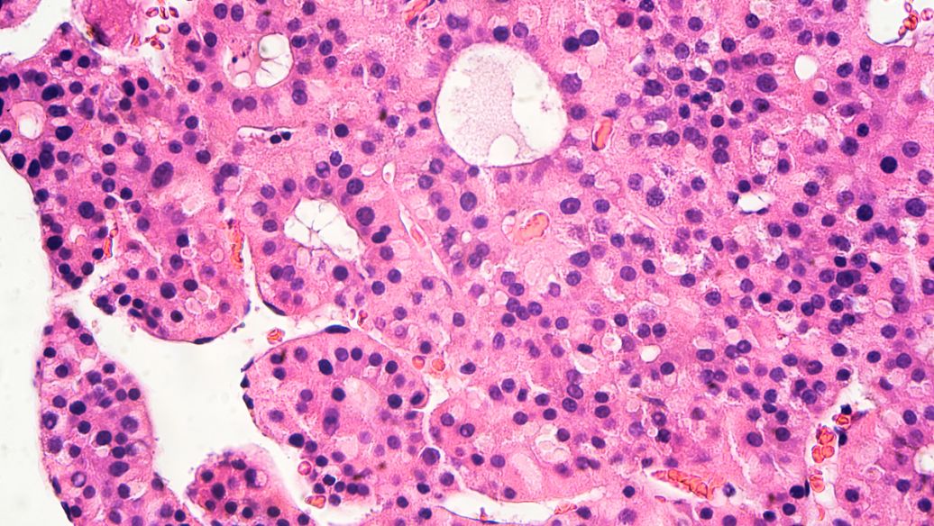
Spinach chloroplast cell under electron microscope
Adjusting Magnification and Focus
Adjusting the magnification levels is essential for observing different aspects of chloroplast ultrastructure. You can start with low magnification to get an overview of the cell architecture and then increase the magnification to focus on specific details of the chloroplasts.
Focusing techniques specific to biological specimens are necessary to achieve optimal resolution of chloroplast membranes while minimizing beam damage. You should use the microscope’s focusing controls to adjust the image and achieve the desired level of detail.
“The quality of the image obtained from an electron microscope depends not only on the instrument itself but also on the skill of the operator.”
By following these steps and mastering the operation of the electron microscope, you can achieve high-quality images of chloroplasts and gain valuable insights into their ultrastructure.
Observing Chloroplast Ultrastructure
Under the electron microscope, chloroplasts display a fascinating ultrastructure that is vital for photosynthesis. As we explore the intricacies of chloroplast structure, we gain insights into the complex processes that occur within these organelles.
Identifying the Chloroplast Envelope
The chloroplast envelope is a double membrane structure that encloses the chloroplast. It consists of two distinct membrane layers: the outer and inner envelope membranes. The space between these membranes is known as the intermembrane space. To identify the chloroplast envelope, look for this characteristic double membrane structure under the electron microscope.
The chloroplast envelope plays a crucial role in regulating the exchange of materials between the chloroplast and the cytoplasm. It is selectively permeable, allowing certain substances to pass through while keeping others out. Understanding the structure and function of the chloroplast envelope is essential for comprehending chloroplast biology.
Examining Thylakoid Membranes and Grana
Thylakoid membranes are the internal membrane system of chloroplasts where light-dependent reactions occur. These membranes are organized into grana stacks, which appear as densely packed, coin-like structures under the electron microscope. Grana are composed of stacked thylakoid discs, and they are the sites where light energy is absorbed and converted into chemical energy.
To examine thylakoid membranes and grana, focus on the regions within the chloroplast where these structures are densely packed. The arrangement of thylakoid membranes and grana can vary depending on the plant species and environmental conditions.
Locating the Stroma and Stromal Lamellae
The stroma is the protein-rich matrix that fills the space between thylakoid membranes. It contains the enzymes responsible for carbon fixation during the Calvin cycle. Stromal lamellae are unstacked thylakoid membranes that connect different grana stacks within the chloroplast, facilitating the exchange of materials and energy.
To locate the stroma and stromal lamellae, look for the regions between grana stacks and the areas surrounding thylakoid membranes. The stroma appears as a less dense region compared to the thylakoid membranes and grana.
- Identify the chloroplast envelope and its components.
- Examine thylakoid membranes and grana stacks.
- Locate the stroma and stromal lamellae.
By following these steps and understanding the ultrastructure of chloroplasts, you can gain a deeper appreciation for the complex processes that occur within these vital organelles.
Image Capture and Processing Techniques
The process of image capture and processing is a critical step in electron microscopy, enabling researchers to visualize chloroplast structures in detail. We will cover the essential techniques for capturing, storing, and processing high-quality electron microscope images of chloroplast ultrastructure.
Digital Image Acquisition Methods
Digital image acquisition has revolutionized electron microscopy, replacing traditional film-based photography with advanced technologies such as CCD cameras and direct electron detectors. These modern detectors offer improved sensitivity and resolution, allowing for the capture of high-quality images with minimal beam damage.
When acquiring images, you need to consider optimal settings such as exposure time, beam intensity, and frame averaging to maximize the signal-to-noise ratio. For instance, increasing the exposure time can improve image quality, but it may also increase the risk of beam damage. Therefore, it’s essential to strike a balance between these factors to obtain the best possible results.
Software Tools for Image Enhancement
Once images are acquired, various software tools can be employed to enhance and process them. Both commercial packages and open-source alternatives are available, offering a range of features such as contrast adjustment, noise reduction, and sharpening. These tools enable researchers to improve the visibility of chloroplast features and highlight specific structural details.
Advanced processing methods, including image stitching and digital filtering, can also be applied to create large field-of-view montages and emphasize particular structural features of chloroplasts. For example, image stitching can be used to create a comprehensive view of a chloroplast, while digital filtering can help to remove noise and enhance the visibility of thylakoid membranes.
| Software Tool | Features | Application |
|---|---|---|
| ImageJ | Contrast adjustment, noise reduction, sharpening | Enhancing chloroplast features |
| Adobe Photoshop | Image stitching, digital filtering | Creating large field-of-view montages |
| GIMP | Contrast adjustment, noise reduction, sharpening | Enhancing chloroplast features |
By combining advanced image acquisition methods with sophisticated software tools, researchers can gain a deeper understanding of chloroplast ultrastructure and its significance in plant cells.
Analyzing Chloroplast Structure from EM Images
The intricate details of chloroplast structure can be revealed through the analysis of EM images. Electron microscopy provides high-resolution images that allow researchers to examine the morphology of chloroplasts within plant cells. By analyzing these images, scientists can gain insights into the organization and function of chloroplasts, which are essential for photosynthesis.
Measuring Chloroplast Dimensions
Accurate measurement of chloroplast dimensions is crucial for understanding their structure and function. Using calibrated image analysis software, researchers can measure the length, width, and cross-sectional area of chloroplasts. This information can be used to determine the number and distribution of chloroplasts within different cell types, providing insights into cellular specialization.
- Measure the length and width of chloroplasts to determine their size and shape.
- Calculate the cross-sectional area to understand the overall morphology.
- Use these measurements to compare chloroplasts from different plant species or under various environmental conditions.
Quantifying Thylakoid Membrane Arrangements
Thylakoid membranes are critical for photosynthesis, and their arrangement within chloroplasts can be quantified using EM images. Researchers can measure the height and diameter of grana stacks, as well as the number of thylakoids per granum. Additionally, calculating the ratio between grana and stromal thylakoids can provide insights into photosynthetic adaptation to different light conditions.
- Measure the height and diameter of grana stacks to understand their structure.
- Count the number of thylakoids per granum to quantify thylakoid membrane arrangements.
- Calculate the ratio of grana to stromal thylakoids to assess photosynthetic adaptation.
Identifying Structural Variations
Chloroplasts from different plant species, developmental stages, or environmental conditions can exhibit structural variations. By analyzing EM images, researchers can identify these variations and understand their implications for chloroplast function. This information is essential for studies involving mutants or plants under stress conditions, where abnormal chloroplast structures may be present.
To identify structural variations, researchers should compare EM images from different samples and look for differences in chloroplast morphology, thylakoid membrane arrangements, or other relevant features. By doing so, scientists can gain a deeper understanding of how chloroplast structure relates to function and how it adapts to different conditions.
Advanced Techniques for Chloroplast Research
To gain a deeper understanding of chloroplast structure and function, researchers are turning to advanced techniques that go beyond traditional microscopy methods. These cutting-edge approaches enable scientists to explore the intricacies of chloroplasts with unprecedented precision, revealing new insights into their role within plant cells.
Immunoelectron Microscopy for Protein Localization
Immunoelectron microscopy (immuno-EM) is a powerful technique that allows for the precise localization of specific proteins within chloroplast subcompartments. By conjugating antibodies to electron-dense gold particles, researchers can visualize the distribution of target proteins with high resolution. This method is particularly useful for studying the spatial organization of proteins within the chloroplast, providing valuable information on their functional roles.
The process of immunogold labeling involves tagging antibody molecules with gold particles of varying sizes, enabling the simultaneous detection of multiple proteins. This technique is invaluable for understanding the spatial relationships between different proteins within the chloroplast. By using gold particles of different sizes, researchers can distinguish between various proteins, gaining insights into their interactions and functional coordination.
- Immunoelectron microscopy enables precise localization of specific proteins.
- Immunogold labeling allows for the simultaneous detection of multiple proteins.
- Different sized gold particles are used to distinguish between various proteins.
3D Reconstruction of Chloroplast Structure
Another advanced technique used in chloroplast research is 3D reconstruction, which enables the visualization of chloroplast architecture in three dimensions. Electron microscopy techniques such as electron tomography and serial section reconstruction are employed to generate 3D models from 2D electron micrographs. These computational approaches provide unprecedented views of thylakoid membrane organization and other structural features, enhancing our understanding of chloroplast function.
Correlative Light and Electron Microscopy (CLEM) combines the advantages of fluorescence microscopy with the high resolution of electron microscopy. This approach allows researchers to correlate dynamic information from live-cell imaging with the ultrastructural details provided by electron microscopy, offering a more comprehensive understanding of chloroplast structures and function.
By incorporating these advanced techniques, researchers can gain a more nuanced understanding of chloroplast biology, shedding light on the complex interactions within these organelles and their role in plant cells.
Comparative Analysis: Chloroplasts vs. Other Organelles
Using electron microscopy, researchers can conduct a comparative analysis of chloroplasts and other organelles, shedding light on their distinct features and functions within plant cells. This comparison is crucial for understanding the unique characteristics of chloroplasts and their role in photosynthesis.
Distinguishing Chloroplasts from Mitochondria
Chloroplasts and mitochondria are both organelles found in plant cells, but they have distinct structures and functions. While both have double membranes, their internal organization differs significantly. Chloroplasts contain thylakoid membranes arranged in grana, whereas mitochondria have cristae that project from the inner membrane into the matrix.
To differentiate between these two organelles in electron micrographs, look for the presence of thylakoids and grana in chloroplasts, and cristae in mitochondria. Additionally, chloroplasts are generally larger and more variable in shape compared to mitochondria.
Differences Between Chloroplasts and Other Plastids
Chloroplasts are a type of plastid found in plant cells, but there are other types as well, including chromoplasts, amyloplasts, and etioplasts. Each of these plastid types has distinct internal structures and contents that relate to their specialized functions.
For example, amyloplasts contain large starch granules, while chromoplasts are characterized by the presence of pigment crystals. Etioplasts, found in dark-grown plants, contain prolamellar bodies. By examining the internal structures and contents of these plastids using electron microscopy, researchers can identify their types and understand their roles in different plant cells and developmental stages.
In conclusion, the comparative analysis of chloroplasts and other organelles using electron microscopy provides valuable insights into their ultrastructure and function. By understanding the differences between these organelles, researchers can better appreciate the complex organization of plant cells.
Troubleshooting Common Issues
When observing chloroplasts under an electron microscope, several common issues can arise, compromising the quality of the images obtained. We will address these challenges and provide practical solutions to ensure high-quality observations.
Addressing Sample Preparation Problems
Sample preparation is a critical step in electron microscopy. Issues such as poor fixation, incomplete dehydration, and improper embedding can compromise chloroplast ultrastructure in cells.
- Poor Fixation: Glutaraldehyde and osmium tetroxide are commonly used fixatives. Ensure that the fixation time and concentration are optimized to preserve the membrane and proteins within the cell.
- Incomplete Dehydration: Verify that dehydration is complete to prevent artifacts that can obscure cell structures.
- Improper Embedding: Use the correct embedding medium and technique to maintain sample integrity, ensuring that the cell components are preserved.
Resolving Image Quality Issues
Image quality can be affected by several factors, including poor contrast, beam damage, and charging effects, all of which can impact the visualization of cell structures.
- Poor Contrast: Adjust the staining protocol or use alternative stains to enhance contrast, making it easier to observe proteins and other structures within cells.
- Beam Damage: Minimize beam exposure and adjust the beam current to reduce damage to the sample, preserving the integrity of the cell membrane.
- Charging Effects: Coat the sample appropriately to prevent charging, ensuring clear images of cell components.
Overcoming Staining Challenges
Staining is crucial for visualizing chloroplast structures. Challenges include uneven staining, precipitate formation, and overstaining, all of which can affect the quality of the image obtained.
- Uneven Staining: Ensure uniform stain penetration by optimizing the staining protocol for the cell type being observed.
- Precipitate Formation: Filter stains before use to prevent precipitates that can obscure details in cells.
- Overstaining: Monitor staining time and concentration to avoid overstaining, which can lead to a loss of detail in the cell structures. For example, adjusting the staining time can significantly improve image quality.
By addressing these common issues, researchers can improve the quality of their electron microscopy images, gaining a clearer understanding of chloroplast ultrastructure in cells.
Interpreting Research Results
To gain meaningful insights from electron microscopy studies of chloroplasts, researchers must interpret the results within the context of cellular biology and photosynthesis. The resulting EM images provide critical information about the structural basis of cell function and cellular disease. As we delve into the interpretation of these results, we will guide you through the process of connecting observed structural features of chloroplasts to their functional significance.
Connecting Structure to Function
The ultrastructure of chloroplasts, as revealed by electron microscopy, is intricately linked to their function in photosynthesis and plant metabolism. Variations in thylakoid membrane organization, for example, can significantly impact light harvesting efficiency and energy transfer in different plant species and growth conditions. By examining the structural components of chloroplasts, such as the envelope, thylakoids, and stroma, researchers can infer their functional roles. “The structural organization of thylakoid membranes is crucial for the efficient capture of light energy and its conversion into chemical energy,” as noted in recent studies.
Furthermore, the chloroplast envelope structure plays a critical role in metabolite transport and communication with other cellular compartments. Understanding these structural features is essential for elucidating the functional significance of chloroplasts in plant cells. For instance, the presence of specific proteins within the chloroplast envelope can facilitate the transport of molecules necessary for photosynthesis.
Comparing Results with Published Literature
When interpreting your chloroplast ultrastructure observations, it’s essential to compare them with published literature to identify consistencies and novel findings. By referencing established studies, you can validate your results and place them within the broader scientific context. This involves using reference images and descriptions from published research to support your interpretations.
For example, comparing your observations of thylakoid membrane arrangements with those reported in the literature can help you recognize and interpret structural changes in response to environmental factors or genetic modifications. As one study noted, “The organization of thylakoid membranes in chloroplasts is highly dynamic and responsive to changes in light intensity.” Such comparisons not only enhance the validity of your research but also contribute to a deeper understanding of chloroplast biology.
Conclusion
In conclusion, our comprehensive guide has demonstrated the crucial role of electron microscopy in studying chloroplast ultrastructure within plant cells. Throughout this article, we have explored the essential techniques and methodologies for observing chloroplasts under an electron microscope, highlighting the significance of these organelles in plant cell biology.
The limited resolution of light microscopy necessitates the use of more powerful techniques, particularly electron microscopy, to analyze the details of cell structure. By employing electron microscopy, researchers can gain a deeper understanding of chloroplast structure and its relationship to function, shedding light on the intricacies of photosynthesis.
Our discussion has covered various aspects, from sample preparation to image analysis, emphasizing the importance of careful technique and methodical approach in electron microscopy. As we move forward, emerging trends and advanced techniques in microscopy will continue to reveal new insights into chloroplast function and its role in broader fields such as agriculture, biotechnology, and environmental science.
In summary, the knowledge gained from studying chloroplast ultrastructure using electron microscopy contributes significantly to our understanding of plant cells and their biological processes. We encourage researchers to apply the techniques described in this guide to further explore the fascinating world of cell biology.
References and further readings:
1.Jin, X., Jiang, Z., Zhang, K., Wang, P., Cao, X., Yue, N., … & Zhou, Y. (2018).
Three-dimensional analysis of chloroplast structures associated with virus infection.
Plant Physiology, 176(1), 282–294.
2.Shimoni, E., Rav-Hon, O., Ohad, I., Brumfeld, V., & Reich, Z. (2005).
Three-dimensional organization of higher-plant chloroplast thylakoid membranes revealed by electron tomography.
The Plant Cell, 17(9), 2580–2586.
3.Vesk, M., Dibbayawan, T. P., Vesk, P. A., & Egan, E. A. (2000).
Field emission scanning electron microscopy of plant cells.
Protoplasma, 212(1–2), 10–23.4.Kirchhoff, H. (2019).
Chloroplast ultrastructure in plants.
New Phytologist, 223(2), 565–570.5.Staehelin, L. A. (2003).
Chloroplast structure: from chlorophyll granules to supra-molecular architecture of thylakoid membranes.
Photosynthesis Research, 76(1–3), 185–196.
FAQ
What is the primary function of chloroplasts in plant cells?
Chloroplasts are organelles found in plant cells responsible for photosynthesis, the process of converting light energy into chemical energy in the form of glucose.
How do electron microscopes differ from light microscopes in terms of resolution and magnification?
Electron microscopes use a beam of electrons to produce an image, offering higher resolution and magnification capabilities compared to light microscopes, which rely on visible light.
What are the key components of a chloroplast’s ultrastructure?
The main components include the chloroplast envelope, thylakoid membranes, grana, stroma, and stromal lamellae, all of which play crucial roles in photosynthesis.
What is the significance of TEM and SEM in chloroplast research?
Transmission Electron Microscopy (TEM) and Scanning Electron Microscopy (SEM) are both essential tools for studying chloroplast ultrastructure, with TEM providing detailed internal structures and SEM offering insights into surface morphology.
How do you prepare plant samples for observation with an electron microscope?
Sample preparation involves chemical fixation, dehydration, embedding, sectioning, and staining to preserve the ultrastructure and enhance contrast for clear visualization.
What safety precautions should be taken when working with electron microscopes and related equipment?
Safety measures include wearing protective gear, following proper handling procedures for chemicals and equipment, and ensuring the microscope is correctly maintained and operated.
How can image processing software enhance the quality of electron microscopy images?
Image processing software can improve image contrast, remove noise, and correct for distortions, thereby enhancing the overall quality and usefulness of the images for research purposes.
What are some common issues encountered during sample preparation for TEM, and how can they be addressed?
Common issues include inadequate fixation, incomplete dehydration, and sectioning artifacts. These can be mitigated by optimizing fixation protocols, ensuring thorough dehydration, and using precise sectioning techniques.
Leo Bios
Hello, I’m Leo Bios. As an assistant lecturer, I teach cellular and
molecular biology to undergraduates at a regional US Midwest university. I started as a research tech in
a biotech startup over a decade ago, working on molecular diagnostic tools. This practical experience
fuels my teaching and writing, keeping me engaged in biology’s evolution.


Leave a Comment
Your email address will not be published. Required fields are marked *