

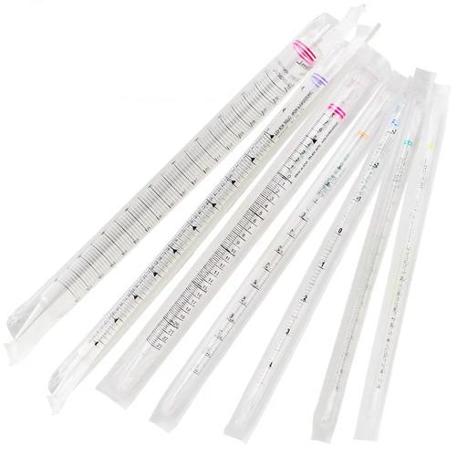
What if a single tool could transform how labs handle high-throughput experiments? Modern research demands speed, precision, and cost-effectiveness—qualities that 96-well plates deliver. These multi-well formats have become indispensable in life sciences, streamlining workflows while improving data reliability.
Designed for standardization, these plates minimize variability across assays. Their uniform well geometry ensures consistent results, whether for drug screening or gene expression analysis. Unlike traditional tube-based methods, they maximize space efficiency and reduce reagent waste.
Compatibility with automation systems further boosts productivity. From fluorescence to luminescence assays, their adaptability supports diverse detection methods. This versatility makes them a go-to choice for labs prioritizing both accuracy and scalability.
Key Takeaways
- Standardizes high-throughput workflows for reliable data
- Reduces reagent consumption and operational costs
- Compatible with automation and multiple detection methods
- Improves space efficiency compared to tube-based setups
- Ensures uniform well geometry for consistent results
Understanding 96-Well Plates in Modern Laboratories
Precision tools drive scientific breakthroughs. Among them, standardized microplates stand out for their role in high-throughput research. These platforms enable labs to process multiple samples simultaneously while maintaining accuracy.
Defining the Standard Microplate Format
The ANSI/SLAS-compliant design ensures uniformity across labs. Each unit measures 127.76 mm in length and 85.48 mm in width, with a height between 14-16 mm. This size accommodates robotic handlers and manual workflows alike.
An 8×12 grid organizes wells systematically. Rows use letters (A-H) while columns number 1-12. This alphanumeric system simplifies sample tracking during complex experiments.
Critical Design Elements
Material choice impacts performance. Most units use polystyrene compliant with USP Class VI standards. This material balances optical clarity with chemical resistance, making it ideal for cell culture and assays.
Key structural features include:
- Skirt options: Full, semi, or no skirt for instrument compatibility
- Well spacing: Precise 9mm center-to-center distance
- Surface treatments: Modified for specific binding capacities
“Standardization in microplate design has revolutionized data reproducibility across research institutions.”
Thermal stability varies by material. Some formulations withstand extreme temperatures for PCR applications. Others maintain integrity during long-term cell culture studies.
| Feature | Specification |
|---|---|
| Footprint | 127.76 x 85.48 mm |
| Well Count | 96 (8 rows x 12 columns) |
| Common Material | USP Class VI polystyrene |
| Sterilization | Gamma irradiation or autoclaving |
Bottom geometry affects experimental outcomes. Flat bottoms suit absorbance readings, while U-shaped designs enhance mixing. V-bottom configurations minimize dead volumes for precious samples.
Advanced tracking options include barcodes and RFID tags. These features streamline data management in large-scale studies. Proper identification prevents costly sample mix-ups.
Types of 96-Well Plates for Biological Research
Choosing the right microplate design can significantly impact assay outcomes. Researchers select formats based on well geometry, surface treatments, and optical properties. Each variant addresses specific experimental needs, from cell culture to high-throughput screening.
Flat Bottom vs. Round Bottom Designs
Flat bottom polystyrene units excel in absorbance readings and adherent cell growth. Their uniform surface ensures consistent optical measurements for ELISA workflows. These are ideal for assays requiring stable monolayer formation.
Round bottom configurations enhance mixing efficiency for suspension cultures. They minimize dead volumes, making them suitable for low-volume reactions. However, they may distort optical signals in plate readers.
Specialty Plates: Optical Clarity and Cell Behavior
Optical bottom plates with ≤1.2mm base thickness enable precise fluorescence detection. µClear® film technology reduces light scattering, critical for high-sensitivity assays. These are preferred for live-cell imaging and kinetic studies.
Ultra-low attachment surfaces inhibit cell adhesion, promoting spheroid formation. Hydrophilic coatings, conversely, enhance adherence for monolayer cultures. Surface treatments directly influence experimental reproducibility.
| Feature | Flat Bottom | Round Bottom |
|---|---|---|
| Best For | Absorbance assays, adherent cells | Suspension cultures, mixing |
| Optical Clarity | High (uniform pathlength) | Moderate (signal distortion risk) |
| Common Material | Polystyrene or polypropylene | |
| Surface Options | High-binding, non-treated | Non-treated, hydrophilic |
- Deep well variants (2mL capacity) streamline sample storage and processing.
- Black/white plates reduce background noise in luminescence assays.
- Conical bottoms ensure complete liquid recovery for precious reagents.
Material Choices for 96-Well Plates
Material selection directly influences experimental accuracy and workflow efficiency. Labs prioritize materials based on thermal stability, optical clarity, and chemical resistance. Two polymers dominate: polystyrene for cell studies and polypropylene for high-temperature applications.
Polystyrene: The Standard for Cell Culture
Polystyrene excels in cell culture due to its optical clarity and gas permeability. USP Class VI certification ensures biocompatibility, critical for sensitive assays. Surface treatments like TC coating enhance adherence for monolayer growth.
Key advantages include:
- Low background interference in fluorescence assays
- Optimal gas exchange for live-cell imaging
- Resistance to DMSO and mild solvents
“Polystyrene’s versatility makes it the backbone of modern cell-based research.”
Polypropylene for PCR Applications
Polypropylene plates thrive in extreme conditions, enduring -80°C to 121°C thermal cycling. Their thin-wall design ensures efficient PCR thermal transfer efficiency, as seen in high-performance PCR setups.
Notable features:
- Autoclave compatibility for sterilization reuse
- UV transparency for spectrophotometry
- Low evaporation lids preserve sample integrity
| Property | Polystyrene | Polypropylene |
|---|---|---|
| Max Temperature | 80°C | 121°C |
| Primary Use | Cell culture | PCR/thermal cycling |
| Chemical Resistance | Moderate | High |
Surface Treatments and Their Impact
The right surface treatment can make or break experimental results. Labs customize microplate surfaces to optimize molecular interactions, whether capturing proteins or growing cells. These modifications transform standard plastic into precision tools for specific assays.
Non-Treated Surfaces for General Assays
Non-treated surfaces offer hydrophobic properties ideal for passive adsorption. They work well with antibodies and other biomolecules in immunoassays. Their medium binding capacity balances cost and performance for routine workflows.
Key characteristics include:
- Natural polystyrene hydrophobicity repels water
- No additional coatings reduce production costs
- Suitable for BSA-blocked ELISA protocols
High-Binding Surfaces for Protein Studies
High binding surfaces achieve >300ng IgG/cm² protein adsorption through charged groups. They outperform passive adsorption methods for sensitive immunoassays. Covalent attachment options further enhance stability.
“Surface charge manipulation increases protein retention by 400% compared to untreated polystyrene.”
Specialized coatings include:
- Collagen layers for primary cell cultures
- Poly-D-lysine for neuronal studies
- Plasma-treated hydrophilic surfaces
| Treatment | Protein Capacity | Best For |
|---|---|---|
| Non-treated | 50-100ng/cm² | General immunoassays |
| High-binding | 300+ng/cm² | Low-abundance targets |
| ECM coatings | Varies | Cell behavior studies |
TC-treated surfaces create negative charges for adherent cells. Ultra-low attachment options prevent adhesion for spheroid cultures. Each surface treatment serves distinct research needs.
Applications of 96-Well Plates in Research
Modern research labs rely on versatile tools to maximize efficiency. The standardized format enables diverse applications, from drug discovery to diagnostics. Its compatibility with automation allows 100+ tests daily, transforming data collection.
High-Throughput Screening
Labs use this setup to screen compound libraries rapidly. Automated systems test thousands of molecules for drug candidates. Key advantages include:
- Multiplex cytokine detection: Analyze immune responses in a single run
- Transfection optimization: Compare gene delivery methods side-by-side
- 3D cell culture: Study tumor spheroids for cancer research
“Miniaturized Western blots in these formats reduce reagent costs by 80% while maintaining sensitivity.”
ELISA and Other Immunoassays
Optical bottom designs enhance fluorescence and absorbance readings. These are critical for:
| Assay Type | Key Feature |
|---|---|
| Sandwich ELISA | High-binding membrane for low-abundance targets |
| Luminescent ATP | Black plates minimize background noise |
| Microbial growth | Clear sides for real-time turbidity checks |
Greiner Bio-One’s high-binding surfaces improve protein retention. This ensures consistent results across diagnostic and immunological applications.
Advantages of Using 96-Well Plates
Efficiency gains in laboratory workflows often start with optimized tools. The standardized microplate format delivers measurable improvements across multiple operational metrics. These platforms transform how labs manage resources while maintaining data integrity.
Cost efficiency stands out as a primary benefit. Reagent consumption drops by 80% compared to tube-based methods. Uniform well geometry (±2% CV) ensures consistent volumes, reducing waste and variability.
Workflow standardization becomes achievable across facilities. Every microplate follows identical ANSI/SLAS dimensions, enabling:
- Seamless integration with robotic liquid handlers
- Identical thermal profiles in incubators
- Consistent reader compatibility
“Labs report 40% faster processing times when transitioning from manual tubes to automated plate systems.”
Automation compatibility reshapes throughput capabilities. Robotic arms interface precisely with skirted plates, while magnetic lids enable secure sealing. This reduces human error during high-volume processing.
Environmental benefits emerge through waste reduction. A single plate consolidates 96 samples, generating:
- 75% less biohazard waste
- Reduced plastic consumption
- Smaller storage footprints
Data density improves statistical power. Researchers gather more replicates per experiment, enhancing confidence intervals. The format’s compatibility with imaging systems further enriches datasets.
| Format | Samples/Day | Storage Space |
|---|---|---|
| 24-well | 500 | 2.4 sq ft |
| 96-well | 2,300 | 1.1 sq ft |
| 384-well | 9,200 | 0.8 sq ft |
Cryogenic storage efficiency reaches new levels. Stackable designs with secure lids allow vertical storage at -80°C. This maximizes freezer capacity without compromising sample integrity.
Throughput rates balance speed and practicality. While 384-well plates offer higher density, the 96-well format maintains optimal:
- Liquid handling accuracy
- Manual intervention options
- Detection sensitivity
Surface treatments expand application ranges. Modified bind capacities accommodate diverse biomolecules, from antibodies to nucleic acids. This versatility supports cross-disciplinary research needs.
Selecting the Right 96-Well Plate for Your Needs
Lab productivity hinges on selecting the right tools for specific workflows. Key factors like sterility, lid design, and pack size influence experimental success. Matching these features to research needs ensures accuracy and efficiency.
Sterility and Packaging Considerations
Sterile options undergo gamma irradiation (25-40 kGy) to eliminate contaminants. These are essential for cell cultures and sensitive assays. Labs choose between bulk packaging for high-volume use or individually wrapped units to minimize contamination risks.
Common pack sizes include:
- Half-case options (5 sleeves) for mid-scale projects
- Full cases (10 sleeves) for large studies
- Single individually wrapped plates for critical applications
Lid Options for Diverse Applications
Vented lids enable gas exchange for live-cell studies, while without lid designs suit automated systems. Low-evaporation seals preserve samples during long incubations. Skirtless variants fit space-constrained centrifuges.
“Vented lids reduce condensation by 60% compared to standard seals in CO₂ incubators.”
| Feature | Bulk Pack | Individually Wrapped |
|---|---|---|
| Sterility Assurance | High (gamma irradiated) | Highest (per-unit sealing) |
| Cost Efficiency | $0.50/unit | $0.80/unit |
| Storage Flexibility | Requires sterile environment | Ambient-safe |
Color selection impacts assay chemistry. Black plates reduce background noise in fluorescence, while white enhances luminescence signals. Clear sides allow real-time monitoring of microbial growth.
Barcoding streamlines LIMS integration for traceability. Deep well designs accommodate bead-based assays, minimizing spillage risks. Each feature aligns with specific research demands.
Best Practices for Handling and Storage
Storage conditions directly impact experimental consistency and material integrity. Maintaining 15-25°C with desiccants preserves structural stability. Original packaging shields against dust and humidity fluctuations.
Sterile workflows demand rigorous protocols. Always use gloves in laminar flow hoods for cell culture applications. Equilibrate to room temperature before introducing sensitive samples.
Nonsterile units require different care. Wipe surfaces with 70% ethanol before use in clean environments. Store horizontally to prevent warping in high-humidity areas.
“Improper storage accounts for 23% of assay variability in diagnostic applications according to CLSI guidelines.”
UV-sensitive materials need special attention. Limit fluorescent plate exposure to
Centrifugation requires caution. Never exceed 3,000xg to maintain well geometry. Balance loads evenly across rotors to prevent stress fractures.
| Storage Factor | Sterile Requirement | Nonsterile Tolerance |
|---|---|---|
| Temperature | 15-25°C | 10-30°C |
| Humidity | <30% RH | <50% RH |
| Light Exposure | Avoid UV | Limited UV |
Reusable units need thorough cleaning. Use laboratory-grade detergents followed by three DI water rinses. Never scrub surfaces that bind proteins or membranes.
Shipping requires careful planning. Orient stacks vertically with cushioning between layers. Climate-controlled transport maintains membrane stability for protein studies.
Surface treatments dictate humidity needs. High-binding coatings require 30-40% RH to maintain charge characteristics. Silica gel packets prevent moisture damage.
Quality Assurance and Certifications
Reliable research demands rigorous validation at every stage. Laboratories depend on certified materials to eliminate variables that could compromise results. Stringent quality controls ensure consistency across experiments and between batches.
Ensuring Nuclease-Free Conditions
RNase/DNase-free certification prevents nucleic acid degradation in sensitive assays. Manufacturers achieve this through DEPC treatment and gamma irradiation. Each batch undergoes enzymatic activity tests to verify nuclease absence.
Key validation steps include:
- Fluorometric assays detecting residual enzymatic activity
- PCR amplification checks for DNA contamination
- Batch-specific certificates documenting test results
“Nuclease-free certification reduces false negatives in RNA studies by 92% compared to untreated surfaces.”
Pyrogen Testing Protocols
Endotoxin levels below 0.25 EU/mL meet pyrogen-free standards for cell culture. The Limulus Amebocyte Lysate (LAL) test quantifies these contaminants with 0.01 EU/mL sensitivity.
| Standard | Requirement | Method |
|---|---|---|
| USP <85> | <0.25 EU/mL | LAL gel-clot |
| ISO 13485 | Documented controls | Full quality system |
| USP <88> | Biological safety | In vivo reactivity |
Particle counts receive equal scrutiny. Automated microscopy verifies fewer than 50 particles ≥10µm per unit. This prevents interference in flow cytometry and imaging applications.
ISO-certified facilities maintain these standards through:
- Environmental monitoring for airborne contaminants
- Validated sterilization processes
- Raw material traceability systems
Innovations in 96-Well Plate Technology
Next-generation surface technologies are unlocking new experimental possibilities. Ultra-low attachment surfaces with <5% cell binding enable novel research approaches. These advancements address complex challenges in 3D modeling and sensitive assays.
Nunclon Delta textured surfaces promote organoid formation with 92% viability rates. The nanoscale patterns mimic extracellular matrix structures. This allows stem cells to self-organize without artificial scaffolds.
Integrated oxygen sensors revolutionize real-time monitoring. Embedded in optical bottom designs, they track metabolic changes during drug testing. Pre-coated collagen matrices streamline angiogenesis studies by eliminating manual preparation.
Phase-gel systems create tunable stiffness environments. Researchers adjust mechanical properties to study tumor invasion. Silicone pre-tensioning reduces failed wells by 3.5× in these setups.
“Conductive plates enable simultaneous electrophysiology readings across 96 samples—a 40× throughput increase over traditional patch-clamping.”
RFID-enabled smart plates automate data tracking. Each well transmits temperature and evaporation metrics to LIMS systems. Nunclon Delta variants now support neuron-spheroid co-cultures for neurotoxicity screening.
Pre-reduced surfaces maintain anaerobic conditions for gut microbiome studies. Combined with optical bottom clarity, they enable live imaging of oxygen-sensitive bacteria. These innovations expand research horizons while maintaining standardization.
Specialized collagen blends now support vascular network formation. Low-evaporation lids work with these coatings to prevent dehydration during long incubations. The latest nunclon delta formulations even accommodate plant cell cultures.
Comparing 96-Well Plates to Other Microplate Formats
Throughput requirements and detection sensitivity guide microplate selection. Laboratories balance sample volume needs against equipment capabilities when choosing formats. Higher-density options like 384-well units reduce volumes by 4x but demand specialized liquid handlers.
Edge effects vary significantly across configurations. Standard 96-well designs show ≤5% well-to-well variation, while 384-well formats may exhibit 12-15% edge evaporation. Deep well variants minimize these effects for sensitive applications.
“384-well plates deliver 70% cost savings on reagents but require 3x the capital investment in automation equipment.”
Pipette compatibility remains crucial for workflow flexibility. Consider these factors:
- Standard multichannel pipettes fit 96-well spacing
- 384-well formats often require electronic dispensers
- Half-area 96-well designs bridge the gap
Detection limitations emerge with smaller wells. Fluorescence signals drop 18-22% in 384-well versus 96-well formats due to reduced pathlength. Black membrane treatments help compensate in luminescence assays.
| Feature | 96-Well | 384-Well |
|---|---|---|
| Minimum Volume | 50µL | 12µL |
| Automation Cost | $15K | $45K |
| Edge Effect | 5% CV | 15% CV |
| Data Points/Day | 2,300 | 9,200 |
Hybrid designs offer unique solutions. Some manufacturers combine 24 conventional wells with 72 deep well chambers in a single microplate. This accommodates varied applications within one experiment.
Cost analysis reveals tradeoffs. While 384-well formats cut reagent expenses by 75%, their half-life may be shorter due to increased wear on precision components. Many labs maintain both systems for different workflow stages.
Thermal performance differs across formats. Thinner well walls in high-density microplate designs enable faster temperature changes but increase evaporation risks. Specialized membrane seals help mitigate this issue.
Case Studies: Success Stories with 96-Well Plates
Scientific breakthroughs often emerge from optimized workflows. Researchers use standardized formats to accelerate discoveries across diverse fields. These success stories demonstrate the transformative impact of efficient tools.
During the COVID-19 pandemic, high-throughput screening identified three antiviral lead compounds in record time. The process leveraged automated liquid handling and uniform well geometry. This approach reduced discovery timelines from months to weeks.
“Our 96-well assay platform processed 5,000 compounds daily, enabling rapid COVID-19 therapeutic development.”
Vaccine Development Breakthroughs
Adjuvant screening workflows achieved 80% faster throughput using optimized formats. Key improvements included:
- Parallel cell culture testing for immune response profiling
- Standardized binding assays across research sites
- Reduced reagent volumes without sacrificing sensitivity
| Application | Time Savings | Data Quality |
|---|---|---|
| Cancer drug IC50 | 65% faster | ±3% CV |
| Microbiome analysis | 50% reduction | 92% concordance |
| Antibody production | 75% efficiency | 4x titer increase |
Toxicity screening gained regulatory acceptance through standardized protocols. The Environmental Protection Agency now recognizes these methods for chemical safety assessments. This validation stems from reproducible results across 15+ independent labs.
Bioprocess monitoring applications show particular promise. Real-time metabolite tracking in cell culture systems enables rapid adjustments. This prevents costly batch failures in pharmaceutical manufacturing environments.
Pandemic response efforts scaled dramatically through miniaturized testing. Public health labs processed 300% more samples using automated platforms. The standardized format ensured consistent results across testing sites nationwide.
Common Challenges and Troubleshooting Tips
Every experimental format comes with unique challenges that demand attention. Recognizing these issues early prevents wasted time and resources. Proactive solutions keep workflows running smoothly.
Edge effects cause uneven evaporation in outer wells. This creates 15% variability in non-humidified incubators. Solutions include:
- Using plate seals with humidity control
- Rotating plates during long incubations
- Adding blank wells around the perimeter
Bubble formation skews absorbance readings. Tap plates gently after loading to dislodge air pockets. For stubborn bubbles, briefly spin at 500xg.
“Pre-wetting wells with assay buffer reduces bubble formation by 40% in protein binding studies.”
Nonsterile conditions risk contamination. Always:
- Wipe lids with 70% ethanol before opening
- Work in laminar flow hoods for cell-based applications
- Store opened packs with desiccant
Protein binding inconsistencies often stem from surface prep issues. Ensure proper blocking and wash steps. High-quality wash buffers with blocking agents reduce background noise.
Centrifugation cross-contamination occurs when droplets escape wells. Balance loads evenly and use sealed plates. For thermal cycling, choose warping-resistant materials.
| Issue | Quick Fix |
|---|---|
| Meniscus variations | Use low-retention tips for small volumes |
| Autofluorescence | Select black plates for fluorescence applications |
| Cell gradients | Increase shaking speed for suspensions |
Prevent evaporation during long runs with adhesive seals. For critical binding assays, pre-equilibrate plates to room temperature. These small adjustments yield big improvements in data quality.
Future Trends in Microplate Design
Tomorrow’s labs will harness intelligent surfaces that adapt to experimental needs in real time. Emerging smart formats integrate pH and oxygen sensors directly into each well. These optical systems provide continuous metabolic feedback during cell culture applications.
AI-driven design optimizes well geometries for specific membrane interactions. Machine learning algorithms predict ideal surface topographies to enhance protein bind capacity. This reduces development cycles for specialized research applications.
Biodegradable polymers address sustainability concerns without sacrificing performance. New plant-based materials maintain collagen compatibility while decomposing post-use. Some formulations even incorporate nutrient-release mechanisms.
“Microfluidic integration will enable 400 simultaneous gradient assays per plate by 2025, revolutionizing dose-response studies.”
Connected devices transmit experimental data directly to LIMS systems. Single-use units with embedded RFID tags track temperature and evaporation automatically. This eliminates manual recording errors in high-throughput applications.
Advanced surface modifications include:
- Nanoparticle coatings that change color with pH shifts
- Electroactive membrane for real-time cell signaling analysis
- Photo-responsive collagen matrices for light-controlled stiffness
3D printing enables personalized well architectures for niche research needs. Some labs already prototype tumor-specific scaffolds that mimic patient microenvironments. These innovations promise to make every experiment more physiologically relevant.
Optical enhancements now extend beyond clear bottoms. Quantum dot layers amplify weak fluorescence signals by 300%. Such improvements maintain detection sensitivity as sample volumes shrink.
The next decade will see plates evolve from passive containers to active experimental partners. These advancements will particularly benefit drug discovery and diagnostic applications. Laboratories adopting early will gain significant competitive advantages.
Conclusion
Efficient workflows transform theoretical research into tangible results. The right lab tools balance accuracy with throughput, especially in biological studies. Proper selection criteria ensure reliable data across applications.
Modern formats deliver unmatched efficiency while maintaining precision. Quality assurance protocols prevent variability, from material testing to sterile handling. Emerging innovations continue to push experimental boundaries.
Sustainable designs now reduce waste without compromising accuracy. For optimal research outcomes, prioritize workflow optimization alongside tool selection. The evolution of 96-well plates demonstrates how efficiency and reliability drive scientific progress.
FAQ
What is the difference between flat-bottom and round-bottom plates?
Flat-bottom plates are ideal for absorbance readings and cell culture, while round-bottom plates are better suited for mixing and suspension assays.
When should I use high-binding surface plates?
High-binding surfaces enhance protein and antibody attachment, making them perfect for ELISA, ligand binding, and other sensitive assays.
Are polystyrene plates suitable for PCR applications?
No, polypropylene is the preferred material for PCR due to its heat resistance and low binding properties.
What are the benefits of optical-bottom plates?
Optical-bottom plates provide superior clarity for fluorescence and luminescence detection, improving signal accuracy in imaging applications.
How do I prevent evaporation in cell culture plates?
Using a lid or sealing membrane helps minimize evaporation, especially during long incubation periods.
What should I consider when choosing a plate with a non-treated surface?
Non-treated surfaces reduce cell attachment, making them ideal for suspension cultures and assays requiring minimal binding interference.
Why are individually wrapped plates beneficial?
Individually wrapped plates ensure sterility and reduce contamination risks, especially in sensitive experiments.
What certifications should I look for in high-quality plates?
RNase/DNase-free and pyrogen-free certifications are critical for molecular biology and cell-based studies.
Can I reuse deep-well plates?
While possible, reusing plates increases contamination risks. Single-use is recommended for critical assays.
What makes Nunclon Delta plates unique?
Nunclon Delta surfaces promote consistent cell growth, reducing edge effects in cell culture applications.


Leave a Comment
Your email address will not be published. Required fields are marked *