
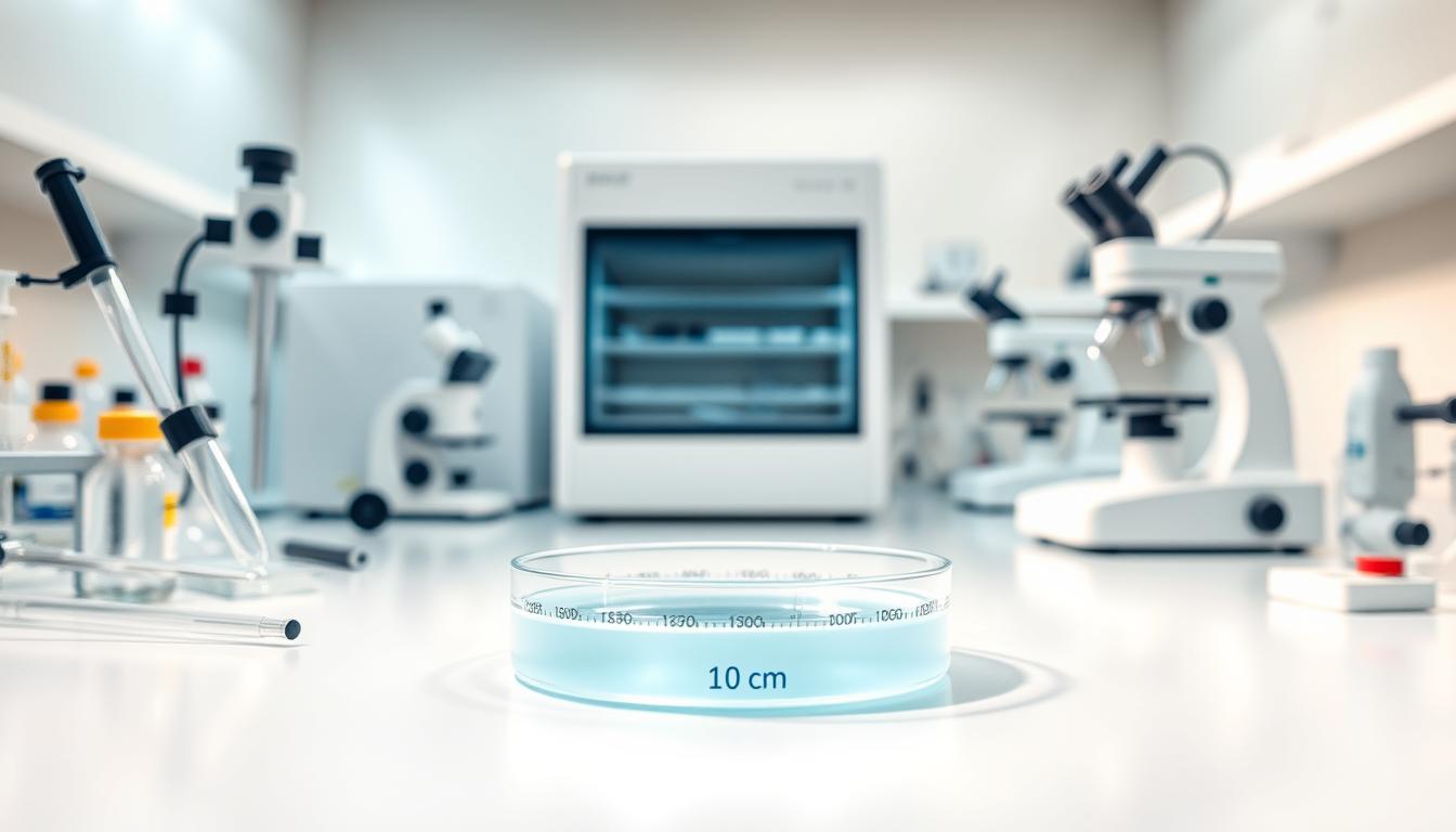
Have you ever wondered what separates groundbreaking scientific research from routine laboratory work in cell culture techniques? The 10 cm dish cell culture method represents a critical foundation for biological investigations, offering researchers a precise and controlled environment for cellular growth and experimentation.
Cell culture dishes provide an essential platform for scientific discovery, enabling researchers to study cellular behavior, develop medical treatments, and explore fundamental biological processes. These specialized 10 cm dish cell culture vessels offer a standardized approach to maintaining and manipulating cells with exceptional precision and reliability.
Mastering cell culture techniques requires a comprehensive understanding of best practices, sterile conditions, and meticulous attention to detail. Scientists must create an optimal environment that supports cell growth while minimizing contamination risks and preserving cellular integrity.
Key Takeaways
- 10 cm dish cell culture provides a standardized method for cellular research
- Precise environmental control is crucial for successful cell cultivation
- Sterile techniques are fundamental to preventing contamination
- Proper cell culture techniques enable breakthrough scientific discoveries
- Understanding cellular growth dynamics is essential for experimental success
Introduction to 10 cm Dish Cell Culture
Cell culture research relies heavily on specialized tissue culture supplies that enable precise biological investigations. The 10 cm dish represents a critical piece of cell culture labware used across numerous scientific disciplines. Its standardized design and optimal dimensions make it an essential tool for researchers studying cell behavior, growth, and interactions.
Researchers select 10 cm dishes for their consistent performance and versatility in biological experiments. These specialized cell culture labware items provide an ideal environment for multiple cell types, supporting critical research objectives.
Importance in Biological Research
The significance of 10 cm dishes in biological research cannot be overstated. These tissue culture supplies offer unique advantages:
- Consistent cell growth surface
- Precise control of cellular environment
- Optimal space for cell expansion
- Easy observation and manipulation
Overview of Applications
Scientists utilize 10 cm dishes across diverse research domains, including:
- Cancer research
- Stem cell studies
- Drug development
- Genetic engineering
Key Considerations
Selecting appropriate 10 cm dish configurations requires understanding specific research parameters. The following characteristics are critical:
| Parameter | Specification |
|---|---|
| Surface Area | 56.7 cm² |
| Seeding Density | 2.2 x 10⁶ cells |
| Maximum Cell Confluency | 8.8 x 10⁶ cells |
| Growth Medium Volume | 12 mL |
Understanding these technical specifications ensures researchers can optimize their experimental conditions and achieve reproducible results in cell culture investigations.
Materials and Equipment Needed
Successful cell culture experiments depend on selecting the right cell biology equipment and sterile cell culture plastics. Researchers must carefully choose their materials to ensure optimal cell growth and experimental integrity.
Proper preparation involves gathering essential supplies that maintain a controlled and contamination-free environment. The following key components are critical for effective cell culture work:
Essential Culture Supplies
- 10 cm tissue culture dishes with specific surface characteristics
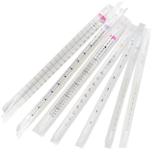
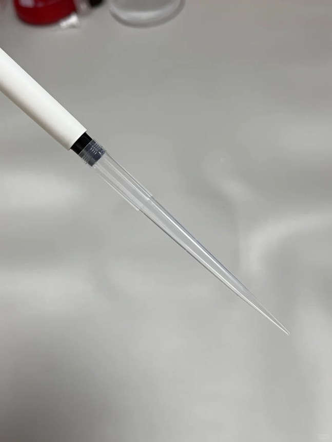
- Sterile pipettes and pipette tips
- Cell culture media
- Specialized cell biology equipment
- Disposable gloves
Recommended Sterilization Techniques
Sterilization is paramount in preventing contamination during cell culture experiments. Proper techniques protect cell integrity and research outcomes.
| Sterilization Method | Application |
|---|---|
| Gamma Irradiation | Sterilizing sterile cell culture plastics |
| Autoclave | Sterilizing reusable glass equipment |
| 70% Ethanol | Surface decontamination |
Safety Equipment
Researchers must prioritize personal and environmental protection when working with cell cultures. Key safety equipment includes:
- Biosafety cabinet
- Personal protective equipment (PPE)
- Biohazard waste containers
- First aid kit
Selecting high-quality cell biology equipment and sterile cell culture plastics creates a foundation for reliable and reproducible research results.
Cell Types Suitable for 10 cm Dish Culture
Cell line propagation is a critical process in biological research, with 10 cm dishes serving as versatile platforms for cultivating diverse cell types. Researchers must understand the specific requirements for different cellular systems to optimize their experimental outcomes.
Adherent cell culture represents a fundamental approach in cellular research, where cells attach and grow directly on the culture dish surface. This method supports numerous cell lines with distinct characteristics and research applications.
Cell Classification for Culture
Two primary cell types dominate 10 cm dish culture techniques:
- Adherent Cells: Grow attached to surface
- Fibroblasts
- Epithelial cells
- Endothelial cells
- Suspension Cells: Float in culture media
- Lymphocytes
- Stem cells
- Blood-derived cells
Common Cell Lines in Research
Several widely used cell lines demonstrate remarkable versatility in 10 cm dish cultures:
- HeLa (cervical cancer cells)
- NIH 3T3 (mouse fibroblasts)
- CHO (Chinese hamster ovary cells)
- HEK 293 (human embryonic kidney cells)
Specialized Cell Type Considerations
Certain specialized cells require meticulous culture conditions. Researchers must carefully select surface coatings, media compositions, and environmental parameters to support optimal cell line propagation and maintain cellular integrity.
Preparing the 10 cm Dish for Cell Culture
Successful cell culture begins with meticulous dish preparation. The 10 cm dish represents a critical platform for monolayer cell growth, providing researchers with an optimal cell culture surface area for experimental studies.
Proper surface preparation directly impacts cell attachment and proliferation. Researchers must carefully consider multiple factors to ensure optimal cell culture conditions.
Surface Coating Options
Effective surface coating enhances cell adhesion and supports monolayer cell growth. Different cell types require specific coating strategies:
- Collagen coating for epithelial cells
- Poly-L-lysine for neuronal cultures
- Fibronectin for endothelial cells
- Laminin for muscle cell lines
Media Selection Tips
Selecting appropriate culture media is crucial for maintaining cell health and promoting robust growth. Consider the following guidelines:
- Match media to specific cell type requirements
- Verify nutrient composition
- Check pH and osmolarity
- Use sterile, high-quality formulations
Sterile Technique Practices
Maintaining aseptic conditions prevents contamination and ensures experimental reliability. Key practices include:
- Use laminar flow hood
- Sterilize all equipment
- Wear appropriate personal protective equipment
- Minimize exposure to external environments
| Parameter | Specification |
|---|---|
| Dish Diameter | 100 mm (10 cm) |
| Growth Area | 56.7 cm² |
| Seeding Density | 2.2 x 10⁶ cells |
| Growth Medium Volume | 12 mL |
Careful preparation of 10 cm dishes creates an ideal environment for cell culture, supporting consistent and reproducible research outcomes.
Inoculation Techniques
Successful cell culture depends on precise inoculation techniques. Researchers must carefully approach the process of introducing cells into cell culture dishes to ensure optimal growth and experimental outcomes. Understanding the nuanced methods of cell distribution and seeding is critical for maintaining cell health and research integrity.
Seeding Density Recommendations
Proper seeding density varies significantly between different cell types. Researchers must consider several key factors when determining the optimal cell concentration:
- Cell line characteristics
- Experimental objectives
- Growth rate of specific cell types
- Desired confluence percentage
Methods for Cell Distribution
Achieving uniform cell distribution in cell biology equipment requires careful technique. Researchers can utilize multiple approaches to ensure even cell spread across culture dishes:
- Gentle pipetting techniques
- Circular swirling motion
- Glass bead method for uniform distribution
- Careful tilting and rotating of culture dishes
| Cell Type | Recommended Seeding Density | Distribution Technique |
|---|---|---|
| Adherent Mammalian Cells | 5,000-10,000 cells/cm² | Gentle pipette distribution |
| Suspension Cells | 1-2 × 10⁵ cells/mL | Circular swirling |
Monitoring Cell Attachment
Successful inoculation requires careful observation of initial cell attachment. Microscopic examination within 24 hours helps researchers assess cell health and distribution. Key indicators include:
- Uniform cell spread
- Typical morphological characteristics
- Absence of debris or contamination
- Consistent cell adherence
Precision in cell culture techniques determines the success of scientific research.
Incubation Conditions
Successful cell culture depends critically on maintaining precise environmental conditions. Researchers using cell culture labware must carefully control several key parameters to ensure optimal cell growth and experimental reliability.
The incubation environment plays a crucial role in cell survival and proliferation. Tissue culture supplies must be selected to support consistent and reproducible conditions across different experimental protocols.
Temperature Optimization
Most mammalian cell cultures require a standard temperature of 37°C. This precise temperature mimics human body conditions and supports optimal cellular metabolism. Variations can significantly impact cell health and experimental outcomes.
- Maintain consistent 37°C temperature
- Use precise digital temperature controls
- Calibrate incubator regularly
CO2 Level Management
Carbon dioxide levels typically range between 5-7%, which helps maintain appropriate pH in culture media. Proper CO2 regulation is essential for cell culture labware performance and cellular stability.
Humidity Control
Relative humidity should be maintained at 95-100% to prevent media evaporation and ensure consistent nutrient availability. Specialized tissue culture supplies include humidity-controlled incubation chambers.
Culture Duration Considerations
Culture duration varies depending on cell type and research objectives. Typical durations range from 24-72 hours, with careful monitoring of cellular growth and morphology throughout the process.
Precision in environmental control determines the success of cell culture experiments.
Monitoring Cell Growth and Health
Successful cell line propagation requires consistent and careful monitoring of cellular health and growth. Researchers must develop keen observational skills to ensure the integrity of their adherent cell culture throughout experimental processes.
Visual inspection serves as the primary method for assessing cell culture conditions. Microscopic examination allows researchers to evaluate critical aspects of cell health:
- Cell morphology and shape
- Density distribution
- Attachment characteristics
- Signs of stress or damage
Techniques for Precise Cell Proliferation Measurement
Quantifying cell growth involves multiple sophisticated approaches. Researchers can utilize various methods to track cell proliferation:
- Manual hemocytometer cell counting
- Automated cell counters
- Metabolic assays
- Fluorescent viability staining
Identifying Potential Contamination
Contamination represents a significant risk in cell culture environments. Researchers must remain vigilant in detecting:
- Bacterial indicators
- Fungal growth patterns
- Mycoplasma presence
- Unexpected color or pH changes in growth media
“Consistent monitoring is the cornerstone of maintaining high-quality cell cultures.” – Cell Biology Research Institute
The recommended monitoring frequency for 10 cm dishes involves daily visual checks and periodic detailed assessments. Tracking key metrics such as cell density (2.2 x 10⁶ cells initial seeding, 8.8 x 10⁶ cells at confluency) ensures optimal experimental conditions for cell line propagation.
Harvesting Cells from a 10 cm Dish
Cell harvesting represents a critical step in cell culture research, requiring precision and careful technique to maintain cell viability and integrity. Researchers working with cell culture dishes must understand the nuanced approaches to extracting cells effectively from sterile cell culture plastics.
Successful cell harvesting involves multiple strategic steps designed to minimize cellular stress and maximize yield. Researchers must select appropriate detachment methods based on specific cell type characteristics and experimental requirements.
Techniques for Cell Detachment
Cell detachment methods can be categorized into two primary approaches:
- Enzymatic detachment using trypsin-EDTA solution
- Non-enzymatic chelating agents like versene
Centrifugation Processes
Proper centrifugation is essential for maintaining cell health during harvesting. Key parameters include:
- Moderate centrifugation speeds (typically 200-300 x g)
- Short centrifugation duration (5-7 minutes)
- Consistent temperature control
Cell Resuspension Methods
The final stage of harvesting involves carefully resuspending cells to create a uniform single-cell suspension. Researchers should use gentle pipetting techniques and appropriate buffers to prevent cellular damage.
Pro tip: Always pre-warm solutions and minimize cell exposure to harsh conditions during the harvesting process.
Troubleshooting Common Issues
Cell culture research demands precision and careful management of cell biology equipment and cell culture labware. Researchers frequently encounter challenges that can compromise experimental results. Understanding common issues and their solutions is crucial for maintaining high-quality cell cultures.
Successful cell culture requires proactive problem identification and strategic intervention. The following sections explore critical troubleshooting strategies for researchers working with 10 cm dishes.
Addressing Contamination Risks
Contamination represents a significant threat to cell culture experiments. Researchers can mitigate risks through rigorous practices:
- Implement strict aseptic techniques
- Use sterile cell culture labware
- Regularly inspect incubation environments
- Maintain clean workspace protocols
Handling Poor Cell Growth
Suboptimal cell growth can result from multiple factors. Key strategies include:
- Verify media quality and composition
- Check cell line viability
- Optimize incubation conditions
- Assess seeding density
Solutions for Abnormal Morphologies
Cell morphology provides critical insights into cellular health. Researchers should investigate potential causes when observing irregular cell shapes or behaviors.
| Morphology Issue | Potential Cause | Recommended Action |
|---|---|---|
| Rounded cells | Nutrient deficiency | Media replacement |
| Detached cells | Enzymatic stress | Adjust trypsinization protocol |
| Irregular growth | Contamination | Sterilize cell biology equipment |
Proactive monitoring and systematic troubleshooting are essential for maintaining robust cell culture experiments.
Documentation and Data Recording
Precise documentation serves as the backbone of successful cell line propagation. Researchers must develop a systematic approach to recording critical information throughout the cell culture process. Maintaining comprehensive records enables scientific reproducibility and supports ongoing research developments.
Effective documentation involves tracking multiple key metrics that provide insights into monolayer cell growth patterns and experimental conditions. Researchers should create detailed logs that capture essential information about cell culture experiments.
Critical Metrics for Documentation
- Cell seeding density: 5.0 x 10⁶ cells
- Surface area measurements: 145 cm²
- Cell confluence levels: 20.0 x 10⁶ cells
- Media composition details
- Incubation conditions
- Passage numbers
Recommended Documentation Practices
Implementing robust documentation strategies requires attention to detail and consistent recording practices. Researchers should utilize specialized software tools designed for managing cell culture data efficiently.
- Take phase contrast microscopic images at key stages:
- Immediately post-thaw
- 24 hours after seeding
- 48 hours after seeding
- At 70-80% confluence
- Record observations from microscope examinations
- Document precise volumes:
- Versene: 10 mL
- Trypsin: 10 mL
- Growth medium: 30 mL
Software Tools for Data Management
Modern laboratory management systems offer sophisticated tracking capabilities for cell culture documentation. These digital platforms enable researchers to maintain comprehensive records, track experimental progress, and analyze cell growth patterns with unprecedented precision.
“Accurate documentation is the cornerstone of reproducible scientific research.”
Future Trends in Cell Culture Techniques
The landscape of cell culture technology is rapidly evolving, with innovative approaches transforming how researchers utilize tissue culture supplies. Advanced 10 cm dishes with specialized surfaces like Nunclon Delta and BioLite are pushing the boundaries of cell yield and morphology research. These emerging technologies are creating new possibilities for understanding cellular interactions and optimizing cell culture surface area.
Cutting-edge developments in cell line development are revolutionizing biological research. Gene editing techniques and personalized cell therapies are expanding the potential of cell culture methodologies. Researchers now have access to customized surfaces such as Nunclon Supra, which support xeno-free and low-serum applications, enabling more precise and adaptable experimental designs.
Technological integration is becoming increasingly sophisticated in cell culturing practices. Automation and artificial intelligence are streamlining processes, allowing for high-throughput screening and more efficient data collection. The integration of advanced tissue culture supplies with intelligent screening technologies promises to accelerate scientific discoveries and improve research methodologies.
The future of cell culture techniques looks promising, with continuous innovations enhancing researchers’ capabilities. From improved dish designs to sophisticated screening methods, scientists can expect more robust, reliable, and efficient tools for exploring cellular dynamics and potential therapeutic applications.
FAQ
What are the primary advantages of using 10 cm dish cell culture?
10 cm dishes provide an optimal surface area for adherent cell culture, allowing researchers to propagate cell lines with consistent growth, maximum cell density, and reliable experimental results. They offer versatility across multiple cell types and are compatible with various research applications, from basic scientific investigation to advanced drug discovery processes.
How do I prevent contamination in my cell culture dishes?
Preventing contamination requires strict sterile technique practices, including working in a biosafety cabinet, using sterile cell culture plastics, wearing appropriate personal protective equipment, regularly disinfecting work surfaces, and maintaining clean incubation conditions. Always use aseptic techniques when handling media, cells, and culture supplies.
What factors should I consider when selecting cell types for 10 cm dish culture?
Consider the cell line’s growth characteristics, including adherent vs. suspension properties, specific nutritional requirements, optimal temperature and CO2 conditions, and compatibility with your experimental goals. Different cell types may require specialized media, surface coatings, and unique culture conditions.
How often should I change the cell culture media?
Media replacement frequency depends on cell type, growth rate, and metabolic activity. Typically, researchers change media every 2-3 days for most cell lines. Monitor cell density, pH indicators, and nutrient consumption to determine the optimal media replacement schedule for your specific experiment.
What are the key indicators of healthy cell growth?
Healthy cell cultures demonstrate consistent morphology, uniform cell distribution, appropriate confluence levels, and active proliferation. Use microscopic examination to assess cell shape, assess attachment quality, and monitor growth patterns. Avoid cultures showing signs of stress, irregular morphology, or reduced metabolic activity.
How do I determine the appropriate seeding density?
Seeding density varies based on cell type, experimental objectives, and desired confluence. Generally, researchers seed between 1-5 x 10^4 cells/cm², adjusting based on specific cell line characteristics and growth rates. Consult cell line-specific protocols and optimize through experimental validation.
What safety precautions are necessary when performing cell culture?
Essential safety precautions include wearing personal protective equipment, working in a biosafety cabinet, using proper waste disposal protocols, handling potentially biohazardous materials carefully, maintaining clean work environments, and following institutional biosafety guidelines and standard operating procedures.
How can I troubleshoot poor cell growth in my 10 cm dishes?
Troubleshooting poor cell growth involves systematically evaluating media composition, culture conditions, cell line viability, potential contamination, and environmental factors. Check incubation parameters, verify media quality, inspect for mycoplasma or bacterial infections, and confirm appropriate cell passage techniques.
What documentation is critical during cell culture experiments?
Comprehensive documentation should include cell line details, passage numbers, seeding densities, media compositions, incubation conditions, observation notes, experimental interventions, and any deviations from standard protocols. Maintain detailed records to ensure experimental reproducibility and facilitate future research analysis.
What emerging technologies are transforming cell culture techniques?
Emerging technologies include advanced gene editing techniques, automated cell culture systems, high-throughput screening platforms, artificial intelligence-assisted monitoring, personalized cell therapies, and innovative culture vessel designs that optimize cell growth and experimental outcomes.


Leave a Comment
Your email address will not be published. Required fields are marked *