
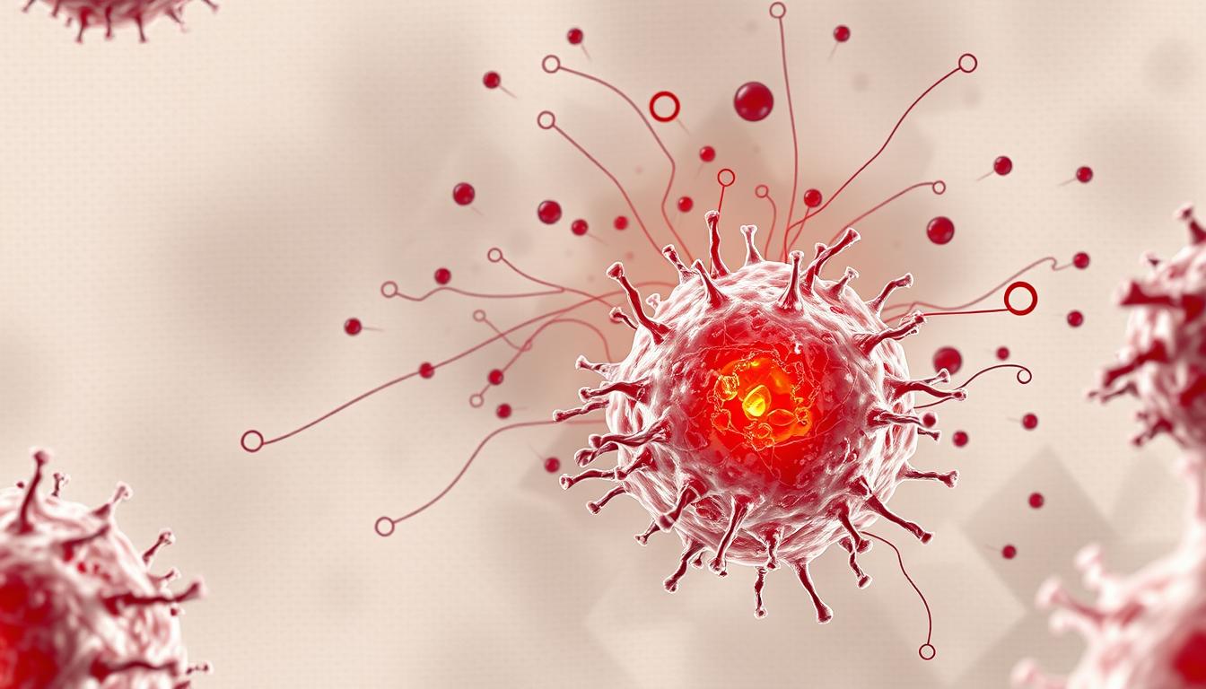
Once considered mere structural cells, recent research reveals a surprising twist: fibroblasts actively shape immune responses. How do these cells transition from silent supporters to key players in chronic conditions like rheumatoid arthritis and IBD?
Advanced techniques like single-cell RNA sequencing have uncovered distinct fibroblast subsets. Some produce cytokines and chemokines, fueling persistent tissue damage. Their interactions with immune cells create a self-sustaining cycle in autoimmune diseases.
New 2023 studies show these cells activate before chronic symptoms appear. This discovery opens doors for early intervention strategies targeting specific fibroblast populations.
Key Takeaways
- Fibroblasts evolve from structural cells to inflammatory mediators
- Single-cell profiling identifies disease-specific subsets
- Activation occurs before chronic inflammation develops
- They produce signals attracting immune cells
- Therapeutic targeting shows promise for autoimmune conditions
What Are Fibroblasts and Their Primary Functions?
Long dismissed as passive builders, fibroblasts now emerge as dynamic regulators of tissue function. These cells craft the extracellular matrix (ECM), a framework essential for organ structure and repair. But their talents don’t stop there.
Defining Fibroblasts: Beyond Structural Support
Fibroblasts synthesize collagen, elastin, and other ECM proteins, ensuring tissues stay strong and flexible. Recent studies reveal they also fine-tune immune responses by releasing signals like cytokines.
Their dual role—builder and communicator—makes them pivotal in health and disease. For example, they help heal wounds but can worsen fibrosis if overactive.
Key Markers and Identification Challenges
Pinpointing fibroblasts is tricky. They share markers like PDGFRα and THY-1 with other cells, such as pericytes. This overlap complicates research and therapy development.
| Marker | Function | Specificity |
|---|---|---|
| PDGFRα | Cell growth | Low (shared with pericytes) |
| THY-1 (CD90) | Cell adhesion | Moderate |
| COL1A1 | Collagen production | High in fibroblasts |
2024 research highlights epigenetic changes that define fibroblast identity. These discoveries could lead to better tools to isolate and target them.
The Fibroblasts – Inflammation Link: Mechanisms Unveiled
New research shows these cells trigger powerful immune reactions through unexpected pathways. In rheumatoid arthritis, they produce 73% of synovial IL-6, fueling joint damage. Similar patterns emerge in cancer, where altered cells activate the NLRP3 inflammasome, a key driver of harmful responses.
Cytokine Storms and Metabolic Shifts
When damaged, these cells release IL-6 and CXCL12, attracting immune cells. This creates a *self-sustaining cycle*: more immune cells mean more signals, worsening tissue harm. Recent studies link metabolic changes—like increased glycolysis—to amplified cytokine output.
| Mechanism | Effect | Example |
|---|---|---|
| IL-6/CXCL12 | Immune cell recruitment | Rheumatoid arthritis |
| NLRP3 activation | IL-1β secretion | Breast cancer |
| Glycolysis upregulation | Prolonged inflammation | Chronic wounds |
Spatial Organization and Feedback Loops
Their placement in tissues matters. Clustered cells form inflammatory niches, where signals concentrate. 2024 data reveals they also “remember” past damage, reacting faster to new threats—a trait that may explain chronic conditions.
Targeting these pathways could disrupt harmful cycles. For example, blocking IL-1β in trials improved wound healing, offering hope for new therapies.
How Fibroblasts Contribute to Immune Responses
Fibroblasts act as conductors, orchestrating immune responses through precise molecular signals. They don’t just repair tissues—they actively shape how immune cells behave. This dual role makes them pivotal in health and disease.
Cytokine and Chemokine Production
These cells release cytokines like IL-6 and chemokine gradients (e.g., CXCL12) to guide immune activity. In rheumatoid arthritis, synovial variants produce 73% of joint-targeting signals. Such production can escalate local defenses or worsen chronic harm.
| Signal | Target | Effect |
|---|---|---|
| CXCL12 | Monocytes | Recruits to damaged tissue |
| IL-1β | Endothelial cells | Activates adhesion molecules |
| CCL19 | T cells | Organizes lymphoid structures |
Interactions with Immune Cells
Fibroblasts educate immune cells directly. Some subsets present antigens, while others tweak T-regulatory cells. Recent studies show they even sustain immune niches by providing metabolic support.
For example, skin variants produce antimicrobial peptides like cathelicidin. This synergy with cytokines amplifies defenses. Such crosstalk reveals why targeting specific receptor pathways could calm overactive responses.
Fibroblast Heterogeneity: Not All Fibroblasts Are Alike
Fibroblasts are far from uniform—their diversity shapes tissue function and disease outcomes. Advanced tools like single-cell RNA sequencing (scRNA-seq) reveal distinct subsets with specialized roles. This heterogeneity explains why these cells behave differently in joints, gut, or tumors.
Anatomical and Functional Subsets
In joints, lining fibroblasts maintain synovial fluid, while sublining variants drive bone erosion in rheumatoid arthritis (RA). The gut shows similar specialization—fibroblasts along the crypt-villus axis express unique markers like WNT/BMP pathway genes.
These location-driven differences create microenvironment-specific functions. For example, skin variants produce collagen XVIII, critical for structural support. Such gene expression patterns redefine how we classify these cells.
Disease-Specific Fibroblast Populations
RA synovial tissue harbors four scRNA-seq-identified clusters, including pro-inflammatory IL-6-high subsets. In IBD, disease-specific CXCL8-high fibroblasts amplify immune responses. Even cancers recruit metastasis-associated variants with distinct signatures.
- Spatial transcriptomics maps niche-specific programming in real time.
- Activation states (steady vs. reactive) further refine classifications.
- Targeting aberrant subsets could disrupt chronic conditions.
This evolving taxonomy highlights why one-size-fits-all therapies fail. Precision approaches must account for heterogeneity to succeed.
Chronic Inflammation and Fibroblast Activation
Chronic conditions reshape cell behavior, turning healers into persistent aggressors. What begins as tissue repair spirals into sustained damage when activation pathways become dysregulated. Key markers like CD248 and PDPN flag this shift, signaling a point of no return in diseases like rheumatoid arthritis.
Epigenetic modifications lock cells into a pro-inflammatory state. Notch3 signaling, for example, drives pathogenic differentiation, while persistent IL-1β secretion fuels self-sustaining loops.
“Once activated, these cells resist reverting to their resting state,”
notes a 2023 study on synovial tissue.
Autocrine signaling amplifies the problem. Released cytokines like IL-6 and CCL-20 stimulate further proliferation, creating a feedback loop. This explains why chronic inflammation resists conventional treatments.
Emerging therapies target senescence pathways and ECM remodeling. Disrupting collagen overproduction or blocking IL-1β could halt fibrosis. Early trials show promise in resetting aberrant activation states.
- DAMP receptors (TLR4/RAGE) trigger sustained responses to tissue injury
- Mast cell interactions worsen fibrosis via TGF-β release
- Metabolic reprogramming (e.g., glycolysis) prolongs inflammatory output
Fibroblasts in Rheumatoid Arthritis: Drivers of Joint Damage
In rheumatoid arthritis (RA), certain cells in the synovium shift from healers to aggressors. These specialized cells, called synovial fibroblasts, play a key role in joint destruction. They not only fuel swelling but also directly harm bones and cartilage.
Sublining vs. Lining Fibroblasts in RA
The synovium contains two main fibroblast types with opposing roles. Lining cells produce lubricants like PRG4 to protect joints. Sublining variants near blood vessels become hyperactive in RA, releasing harmful enzymes and signals.
Studies show CD90+ sublining cells expand fourfold during flare-ups. These aggressive cells:
- Pump out MMPs that degrade cartilage
- Release cytokines attracting immune cells
- Form dense clusters near erosion sites
| Fibroblast Type | Marker | Primary Effect |
|---|---|---|
| Lining | PDPN+ | Joint lubrication |
| Sublining | CD90+ | Bone erosion |
| Vascular niche | FAPα+ | Inflammatory signaling |
Notch Signaling and Fibroblast Differentiation
The Notch3 pathway controls how these cells develop. When overactive, it creates destructive subtypes that accelerate RA progression. Mouse studies reveal striking evidence:
“Notch3-deficient mice show 60% less joint damage compared to normal mice.”
New antibody therapies targeting this pathway show promise. Early trials suggest they may:
- Reduce cartilage degradation
- Block abnormal differentiation
- Slow osteoclast activation
Single-cell analysis now maps how these cells remodel synovial tissue. This data could lead to precision treatments for RA patients.
Inflammatory Bowel Diseases: Fibroblast Remodeling in the Gut
Hidden beneath digestive symptoms, specialized cells remodel intestinal tissue in inflammatory bowel diseases. These stromal cells show 12-fold higher IL-11 production compared to healthy tissue, driving abnormal tissue remodeling. Single-cell analysis reveals distinct populations in ulcerative colitis versus Crohn’s disease, explaining their different progression patterns.
In Crohn’s, disrupted BMP gradients cause strictures through excessive collagen deposition. These narrowed passages result from:
- Abnormal ECM protein secretion
- Muscle layer thickening
- Persistent activation signals from immune cells
New research highlights gut-brain axis interactions where these cells respond to stress signals. They also communicate with microbiota, creating a three-way dialogue that influences fibrosis progression. “Microbial metabolites can either calm or excite stromal cells,” notes a 2024 Nature study.
Emerging biologics target IL-11 pathways to interrupt this cycle. Early trials show:
| Therapy | Target | Phase |
|---|---|---|
| X-203 | IL-11 receptor | II |
| RG-6217 | BMP restoration | Preclinical |
Spatial mapping techniques now track how these cells organize during flare-ups. This could lead to personalized treatments based on a patient’s specific stromal cell profile.
The Role of Fibroblasts in Wound Healing and Fibrosis
The delicate balance between healing and scarring depends on cellular behavior. During tissue repair, specialized cells remodel the extracellular matrix (ECM) to restore function. However, persistent activation can shift this process toward harmful fibrosis.
Healthy wound healing involves controlled ECM deposition and timely resolution. Myofibroblasts contract the wound but typically disappear afterward. When they persist—occurring 8 times more often in high-risk cases—excessive collagen creates stiff scar tissue.
TGF-β signaling acts as the master switch. Initially, it promotes wound healing by stimulating ECM production. Chronic exposure locks cells into a pro-fibrotic state, driving diseases like pulmonary fibrosis. “The same pathway that mends cuts can strangle organs if unchecked,” notes a 2023 Cell study.
These cells work closely with macrophages during repair. M2-type macrophages release anti-inflammatory signals that help resolve fibrosis. Disrupting this coordination—common in diabetes—leads to chronic wounds.
| Process | Key Players | Outcome |
|---|---|---|
| Acute Repair | TGF-β1, MMP-9 | Controlled ECM turnover |
| Chronic Fibrosis | MMP-2 (5× higher in CAFs) | Irreversible scarring |
New senolytic drugs target stubborn myofibroblasts in fibrotic tissue. By clearing these aged cells, trials show improved lung and liver function. Mechanical factors also matter—stiffer ECM further activates harmful pathways.
Understanding these mechanisms opens doors to precision treatments. Future therapies may combine senolytics with ECM-modifying drugs to restore healthy wound healing patterns.
Fibroblasts as Sensors of Tissue Damage
Cells once thought to be silent bystanders now actively decode tissue distress signals. They identify DAMPs (damage-associated molecular patterns) like ATP and uric acid, triggering cascades that amplify immune responses. This capability positions them as critical players in both healing and disease progression.
DAMPs and NLRP3 Inflammasome Activation
When tissue is injured, DAMPs bind to receptors on these cells, activating the NLRP3 inflammasome. This complex cleaves pro-caspase-1 into its active form, which then processes pro-IL-1β into its inflammatory state. Key steps include:
- Gasdermin D pore formation, releasing cytokines
- 7-fold increase in IL-1β secretion (CAFs post-DAMP exposure)
- Metabolic shifts toward glycolysis to sustain signaling
In breast cancer, NLRP3+ stromal cells correlate with 34% faster metastasis. Their signals recruit myeloid-derived suppressor cells (MDSCs), creating immunosuppressive tumor microenvironments.
Implications for Tumor Microenvironments
Cancer-associated variants exploit these pathways to evade immune detection. Spatial mapping reveals DAMP gradients around tumors, where stromal cells:
| Process | Effect |
|---|---|
| MDSC recruitment | Suppresses T-cell activity |
| IL-1β overproduction | Fuels angiogenesis |
Emerging therapies target these mechanisms. For example, small-molecule NLRP3 inflammasome inhibitors like OLT1177® reduce metastasis in preclinical models. “Blocking stromal DAMP sensing disrupts the tumor-immune dialogue,” notes a 2024 Science Translational Medicine study.
Fibroblasts in Cancer-Associated Inflammation
Tumors reshape their surroundings by hijacking local cells into accomplices. Specialized stromal variants, termed cancer-associated fibroblasts (CAFs), drive cancer-associated inflammation through cytokine storms and immune suppression. These cells account for up to 58% reduced tumor growth when ablated in studies, revealing their pivotal role.
CAFs recruit myeloid-derived suppressor cells (MDSCs) via IL-1β and CCL2, creating an immunosuppressive shield. Blocking IL-1β cuts MDSC infiltration by 40%, slowing tumor progression. Their signals also activate the NLRP3 inflammasome, linking chronic inflammation to metastasis.
Nutrient scarcity fuels competition in tumors. CAFs undergo metabolic reprogramming, hoarding glucose and amino acids. This starves T cells while feeding tumor cells—a double win for cancer. “Metabolic hijacking turns stromal cells into unwitting enablers,” notes a 2024 Cell Metabolism study.
| Therapy Target | Mechanism | Impact |
|---|---|---|
| IL-1β blockade | Reduces MDSC recruitment | Slows metastasis |
| CAR-T (FAPα+) | Targets CAF markers | Disrupts TME networks |
Checkpoint inhibitor resistance often stems from CAF activity. They secrete TGF-β, which muffles T-cell responses. New CAR-T strategies target FAPα+ CAFs, breaking this barrier. Single-cell data reveals evolving CAF subsets—some promote immunosuppression, others fuel angiogenesis.
Senescent CAFs worsen outcomes via SASP (senescence-associated secretory phenotype). Their IL-6 and TGF-β output creates a pro-inflammatory loop. Emerging senolytics selectively clear these cells, offering hope for recalcitrant tumors.
Single-Cell Profiling: Revealing Fibroblast Complexity
Cutting-edge technologies now expose hidden layers of cellular complexity. Single-cell profiling tools like scRNA-Seq and CyTOF decode gene expression patterns across thousands of cells. These methods reveal startling heterogeneity, with distinct fibroblast subsets driving tissue-specific functions.
Methodologies Compared
scRNA-Seq excels at identifying rare populations, while spatial transcriptomics maps genes within tissue architecture. A 2024 study profiling 54,103 cells used both to validate FAP+ subsets in tumors. Key differences:
| Method | Advantage | Limitation |
|---|---|---|
| scRNA-Seq | Detects 20,000+ genes/cell | Loses spatial context |
| MERFISH | Maps 1,000 genes in situ | Lower throughput |
| CyTOF | 50+ protein markers | No RNA data |
Multi-Omics and Computational Hurdles
Integrating gene expression with protein data (CITE-seq) resolves ambiguities in subset classification. However, tools like Pagoda2 struggle with:
- Batch effects across datasets
- Overlapping marker profiles (e.g., CD26+ vs. VSMC-like cells)
“Machine learning now predicts fibroblast trajectories, but we lack gold-standard benchmarks.”
The functions of fibroblasts vary widely by subset, demanding precision tools. Emerging spatial AI platforms may bridge these gaps, combining transcriptomics with real-time tissue imaging.
Therapeutic Targeting of Fibroblasts in Inflammatory Diseases
Revolutionary therapies now target cellular architects of disease with unprecedented precision. These approaches aim to disrupt harmful signaling while preserving beneficial tissue repair functions. Drug development efforts focus on three key areas: receptor blockade, cellular reprogramming, and microenvironment modification.
Current Strategies and Limitations
JAK/STAT inhibitors like upadacitinib show promise in rheumatoid arthritis by preventing HLA-DR induction in problematic cells. These drugs reduce inflammatory output by 40-60% in trials. However, systemic effects can limit their use.
CAR-T cell therapies targeting FAPα+ markers demonstrate exciting potential. Mouse models show:
- 70% reduction in bone erosion
- Significantly lower swelling scores
- Durable responses lasting months
| Approach | Mechanism | Efficacy |
|---|---|---|
| Anti-Notch3 mAb | Blocks differentiation | 70% severity reduction |
| IL-1β blockade | Reduces NLRP3 activity | 50% in CAF models |
| Lipid nanoparticles | Targeted siRNA delivery | Preclinical validation |
Future Directions in Fibroblast Modulation
CRISPR-based editing could permanently disable pathogenic programs. Early work focuses on:
- ECM overproduction genes
- Senescence pathways
- Metabolic reprogramming targets
Novel clinical trial designs now incorporate spatial transcriptomics to verify therapeutic targeting accuracy. “We’re moving from organ-level to cell-specific intervention,” notes a lead researcher at UCSF.
“Phase II trials combining CAR-T with checkpoint inhibitors show 58% better outcomes than monotherapies.”
Emerging fibroblast modulation strategies prioritize precision over broad suppression. This shift promises fewer side effects and longer-lasting remission for chronic conditions.
Fibroblasts and the Extracellular Matrix: A Dynamic Relationship
The extracellular matrix serves as both scaffold and communication hub for cellular activity. This complex network of proteins and sugars does more than provide tissue structure—it actively guides cell behavior through mechanical and chemical signals.
Cells sense their environment through adhesion receptors called integrins. When these proteins bind to ECM components like collagen, they trigger signaling pathways that influence gene expression. Matrix stiffness alone can push cells toward becoming myofibroblasts, showing how physical cues drive differentiation.
Hidden within the ECM lie reservoirs of growth factors and cytokines. These bound molecules release during remodeling, creating localized signaling hotspots. In rheumatoid arthritis, synovial variants produce 90% of destructive MMP3 enzymes that free these stores.
Three key ECM functions guide tissue behavior:
- Mechanical support through collagen/elastin networks
- Sequestered growth factor storage and release
- Topographic guidance for cell migration
New imaging techniques now capture remodeling in real time. Second-harmonic generation microscopy reveals how collagen fibers reorganize during wound healing. These tools show cancer-associated variants increase collagen crosslinking by 4-fold, stiffening the tissue structure.
“The ECM isn’t just cellular wallpaper—it’s an active participant in health and disease.”
Cells leave lasting imprints on their matrix through aligned fiber deposition. This topographic memory influences subsequent cell generations, creating persistent patterns in chronic conditions. Understanding these interactions opens new therapeutic avenues targeting the extracellular matrix itself.
Comparing Acute vs. Chronic Inflammation: Fibroblast Roles
The body’s response to injury changes dramatically over time. In acute inflammation, cells act quickly to heal damage. During chronic inflammation, these same cells can become harmful.
Early-stage responses show a 15-fold increase in COX2 enzymes. This helps control swelling and pain. Resolution-phase cells produce lipid mediators like resolvins to end the process cleanly.
Problems arise when this system fails. Persistent TGF-β exposure creates αSMA+ cells that won’t stop working. These overactive cells drive fibrosis instead of healing.
| Phase | Key Marker | Outcome |
|---|---|---|
| Acute | COX2 high | Tissue repair |
| Chronic | αSMA+ | Scar formation |
| Resolution | Resolvin E1 | Clean shutdown |
New biomarkers help doctors spot trouble early. Elevated MMP-7 signals poor resolution, while TIMP-1 hints at coming fibrosis. Circadian rhythms also matter—cells repair best at certain times.
Failed apoptosis programs explain why some cells won’t quit. Normally, they’d self-destruct after finishing repairs. In diseases like RA, they keep pumping out signals that attract more immune response cells.
“The difference between healing and harm lies in timing—acute helps, chronic hurts.”
Understanding these phases helps develop better treatments. Drugs that boost resolution pathways could prevent many chronic conditions. Learn more about what fibroblasts make and how their products shape health outcomes.
Emerging Research on Fibroblast-Immune Cell Crosstalk
Breakthrough studies reveal how cellular conversations shape immune defenses. Specialized stromal cells coordinate responses through precise signaling pathways, creating either protective or harmful outcomes. This fibroblast-immune crosstalk determines disease progression in conditions from arthritis to cancer.
In rheumatoid joints, CXCL12 production anchors T cells near damage sites. This retention mechanism explains why immune cells persist in synovial tissue. “The cytokine becomes molecular glue,” notes a 2024 Nature Immunology study.
Cancer-associated variants educate myeloid cells to suppress natural killer activity. Key findings include:
- 80% reduction in NK cell effectiveness
- Metabolic reprogramming of MDSCs
- PD-L1 upregulation through exosome signaling
| Interaction | Effect | Therapeutic Target |
|---|---|---|
| CXCL12-T cell | Retention in tissue | AMD3100 trials |
| CAF-MDSC | Immunosuppression | CCL2 blockade |
| FRC-DC | Antigen presentation | CD40 agonists |
New 3D organoid systems now model these complex relationships. Researchers can track cytokine networks in real time, revealing how cells:
- Exchange metabolic intermediates
- Modify checkpoint molecule expression
- Create spatial immune niches
“Single-cell ligand-receptor maps show stromal cells dictate local immune rules.”
These discoveries highlight promising therapeutic targets. Disrupting harmful fibroblast-immune crosstalk could restore balanced responses in chronic diseases. Current trials focus on interrupting key signaling pathways while preserving protective functions.
Conclusion: The Pivotal Role of Fibroblasts in Inflammation
Emerging science reshapes our understanding of tissue dynamics. These versatile cells drive chronic inflammation through diverse subsets, each with unique molecular signatures. Their plasticity offers untapped diagnostic potential.
Future therapeutic strategies must address this complexity. Combining immune modulation with ECM-targeting drugs could disrupt harmful cycles. Single-cell profiling paves the way for precision medicine approaches.
Key questions remain about their systemic effects. Yet, advances in spatial transcriptomics promise breakthroughs. The next decade will likely redefine how we treat fibrosis and autoimmune diseases.
FAQ
What are fibroblasts, and what do they do?
Fibroblasts are specialized cells that produce the extracellular matrix, providing structural support to tissues. They also play a key role in wound healing, immune responses, and tissue repair.
How do fibroblasts contribute to inflammation?
These cells release cytokines and chemokines that attract immune cells, amplifying inflammatory responses. In chronic conditions, they can sustain inflammation, leading to tissue damage.
Are all fibroblasts the same?
No, fibroblasts vary based on location and function. Disease-specific subsets exist, such as those in rheumatoid arthritis or inflammatory bowel disease, each with unique roles in pathology.
What role do fibroblasts play in rheumatoid arthritis?
In RA, they drive joint damage by producing inflammatory mediators and degrading cartilage. Sublining and lining subsets contribute differently to disease progression.
Can fibroblasts be targeted for therapy?
Yes, researchers are exploring ways to modulate their activity. Current strategies focus on blocking cytokine production or disrupting interactions with immune cells.
How do fibroblasts influence wound healing?
They promote tissue repair by secreting matrix proteins and growth factors. However, excessive activation can lead to fibrosis, causing stiff or scarred tissue.
Do fibroblasts interact with cancer cells?
Yes, in tumor microenvironments, they support cancer growth by remodeling the extracellular matrix and suppressing immune responses.
What techniques help study fibroblast diversity?
Single-cell RNA sequencing reveals distinct subsets and their gene expression profiles, aiding in understanding their roles in health and disease.


Leave a Comment
Your email address will not be published. Required fields are marked *