


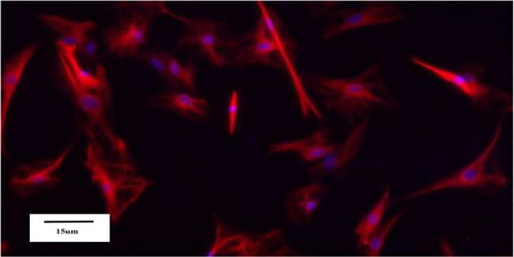
(Image Source:Xiangyu zhai and WeiWang)
The hepatic stellate cells (HSCs) localize at the space of Disse in the liver and have multiple functions. They are identified as the major contributor to hepatic fibrosis. Here, we described protocol for isolation of human HSCs.
Materials and Reagents
All materials must be filtered with a 0.20 μm sterile filter.
1.Enzyme H solution: Added Three milliliters of Dulbecco’s modifed Eagle’s medium (DMEM) was added to the lyophilized Enzyme H powder and mixed them evenly by shaking gently. Aliquots were cryopreserved at −20 ℃. Repeated freezing and thawing were avoided. (Miltenyi Biotec, 130-093-237, Germany)
2.Enzyme R solution: DMEM (2.7 ml) was added to the lyophilized Enzyme R powder. After mixing, the solution aliquots were cryopreserved at −20 ℃. Repeated freezing and thawing were avoided. (Miltenyi Biotec, 130-093-237, Germany)
3.Enzyme A solution: One milliliterl of DMEM was added to the lyophilized Enzyme a powder; vigorous shaking was avoided while mixing. The aliquots were cryopreserved at −20 ℃. (Miltenyi Biotec, 130-093-237, Germany)
4.DNase I (Roche, Switzerland)
5.Nycodenz Separation Solution (density 1.131 g/ml, Beijing Solarbio Science & Technology Co., Ltd.)
6.RPMI 1640 medium (Gibco, USA)
7.DMEM/F12 medium (Gibco, USA)
8.Gey’s balanced salt solution (GBSS, Sigma, USA)
9.Phosphate-bufered saline (PBS, Gibco, USA)
10.Penicillin-streptomycin (Gibco, USA)
11.Fetal bovine serum (FBS, Gibco, USA)
12.Trypan blue (Beyotime Biotechnology, China)
Equipment
1.Standard laboratory equipment
2.Precooled centrifuges (5810R, Eppendorf, Germany)
3.Sterile surgical instruments and preparation tools
4.Sterile 15ml plastic tubes (Ucallm, China)
5.Sterile 50ml plastic tubes (Ucallm, China)
6.Pasteur pipettes (Ucallm, China)
7.Standard cell culture room equipped with a sterile hood
8.Petri dishes(Ucallm, China)
9.70μm cell strainer (Ucallm, China)
10.Cell counting chambers (Biologix, 07–2104, China)
11.Inverted fluorescence microscope (NIKON, Ti-S, Japan)
12.Gentle MACS tissue processor (Miltenyi Biotec, 130-093-235, Germany)
13.Gentle MACS C Tube (Miltenyi Biotec, 130-093-237, Germany)
Procedure
1. Preparation of a single cell suspension from human hepatic tissue
1.1 Surgically removed fresh normal liver tissues were processed and collected in an ice box at 4 ℃ under aseptic conditions.
1.2 The tissues were quickly transported to the cell culture chamber. The specimen was
placed in a Petri dish and washed 5 times with a sterile PBS solution to remove residual blood from the tissue surface.
1.3 The connective tissues were trimmed away as much as possible, and the tissues were cut into blocks approximately 1 cm in diameter. The bile ducts, and blood vessels were trimmed away as much as possible using ophthalmic scissors, and the tissue were blocks were further chopped into small approximately 1-mm pieces and placed into a glass Petri dish.
1.4 The premixed working solution (100μl of penicillin-streptomycin, 250μl of Enzyme H, 150μl of Enzyme R, and 20 μl of Enzyme A) was added to 5 ml of DMEM and mixed well to make the mixed-enzyme solution (required for 2–4 g of tissues).
1.5 To prepare the single cell suspension of human liver tissue, the mixed-enzyme solution was prewarmed in a 37 ℃ water bath for 10 min before adding it to the tissues. The enzymatic treatment was carried out in the GentleMACS™ tissue processor at 37 ℃ for 15 minutes with constant shaking using the h_tumor_01.01 program.
2.Isolation of human hepatic cells
2.1 The enzymatic digestion was stopped by diluting the enzyme solution with 10ml of DMEM containing penicillin-streptomycin and FBS. The solution was filtered through a SmartStrainer filter (70 μm), and the filter was rinsed with DMEM containing penicillin-streptomycin and FBS after cell filtration.
2.2 The filtered cell suspension was centrifuged at 50 g for 7minutes (4 ℃) to isolate the hepatocytes, which were contained in the pellets.
2.3 The supernatant was centrifuged again at 450 g for 8 minutes (4 ℃); then, the supernatant was discarded, and the pellet containing the HSCs was collected.
2.4 The pellet was suspended in 7 ml of DMEM and centrifuged at 450 g for 7 minutes (4 ℃). The supernatant was removed as much as possible. One hundred fifty microliters of DNase solution and 10 ml of 18% Nycodenz were added sequentially to resuspend the pellet.
3. Nycodenz density gradient centrifugation
3.1 Five milliliters of the cell suspension in the 18% Nycodenz solution was added to each of two 15 ml centrifuge tubes. Then, 3 ml of 12% Nycodenz and 3 ml of 8.2% Nycodenz were added gently. After further mixing with 2ml of GBSS, the solutions were subjected to horizontal centrifugation at 1450g for 22min (4 ℃) without braking.
3.2 After the centrifugation was completed, the HSCs were aggregated at the interface of the 8.2% Nycodenz solution and the GBSS solution and the 18% and 12% Nycodenz solutions.
3.3 The cells were carefully collected into another centrifuge tube and then evenly mixed in 4ml of 1×PBS. The solution was centrifuged at 150 g for 8min at 4 ℃. The resulting supernatant was discarded, and the cell pellet was resuspended in RPMI 1640 medium containing FBS, followed by trypan blue staining and cell counting.
References
1.Zhai, X., Wang, W., Dou, D. et al. A novel technique to prepare a single cell suspension of isolated quiescent human hepatic stellate cells. Sci Rep 9, 12757 (2019). https://doi.org/10.1038/s41598-019-49287-7
2.Shang, L., Hosseini, M., Liu, X.et al. Human hepatic stellate cell isolation and characterization. J Gastroenterol 53, 6–17 (2018). https://doi.org/10.1007/s00535-017-1404-4
Manufacturer’s Link
Miltenyi Biotec: https://www.miltenyibiotec.com
Roche: https://www.roche.com
Solarbio: https://www.solarbio.com
Thermo Fisher Scientific: https://www.thermofisher.com ›home ›brands ›gibco
Sigma-Aldrich: https://www.sigmaaldrich.com
Beyotime: https://www.beyotime.com
Eppendorf: https://www.eppendorf.com
biologixlab.com: https://www.biologixlab.com
Nikon USA: https://www.nikonusa.com
Disclaimer:
If any infringement is identified, please contact 【info@ucallmlabs.com】for content removal.
Product Name | Catalog Number | Specification |
Syringe filters | F1121 | 0.22 μm |
1000 μL Tip | L1110 | 100~1000 μL |
Cell strainer | F2040 | 40 μm |
Cell strainer | F2100 | 100 μm |
Petri dishes | C2510 | 100 mm |
Human Primary Cells
The Immune Cell System
Hepatic Cell System
- Human Hepatic Sinusoidal Endothelial Cells
- Human Intrahepatic Biliary Epithelial Cells
- Human Hepatocytes
- Human Hepatic Stellate Cells
- Human Hepatic Macrophages
- Human Gallbladder Fibroblasts
- Human Liver-derived Mesenchymal Stem Cells
Neurons Cell System
- Human Brain Microvascular Endothelial Cells
- Human Brain Vascular Smooth Muscle Cells
- Human Brain Vascular Adventitial Fibroblasts
- Human Brain Vascular Pericytes
- Human Choroid Plexus Endothelial Cells
- Human Choroid Plexus Epithelial Cells
- Human Choroid Plexus Fibroblasts
- Human Meningeal Cells
- Human Leptomeningeal Pericytes
- Human Dural Fibroblasts
- Human Neurons
- Human Cerebellar Granule Cells
- Human Neurons-hippocampal
- Human Oligodendrocyte Precursor Cells
- Human Schwann Cells
Skeletal Cell System
- Human Calvarial Osteoblasts
- Human Osteoblasts-femoral
- Human Chondrocytes-articular
- Human Skeletal Muscle Cells
- Human Skeletal Muscle Myoblasts
- Human Synoviocytes
- Human Nucleus Pulposus Cells
- Human Annulus Fibrosus Cells
- Human Bone Marrow-Derived Mesenchymal Stem Cells
Cardiac Cell System
- Human Cardiac Microvascular Endothelial Cells
- Human Coronary Artery Endothelial Cells
- Human Coronary Artery Smooth Muscle Cells
- Human Aortic Endothelial Cells
- Human Aortic Smooth Muscle Cells
- Human Aortic Adventitial Fibroblasts
- Human Cardiac Myocytes
- Human Cardiac Fibroblasts
- Human Ventricular Cardiac Fibroblasts
- Human Pericardial Fibroblasts
Dermal Cell System
- Human Dermal Microvascular Endothelial Cells
- Human Dermal Lymphatic Endothelial Cells
- Human Dermal Blood Endothelial Cells
- Human Epidermal Keratinocytes
- Human Epidermal Melanocytes
- Human Dermal Fibroblasts
- Human Scalp Fibroblasts-fetal
Adipose Cell System
- Human Adipose Microvascular Endothelial Cells
- Human Preadipocytes
- Human Adipose-derived Mesenchymal Stem Cells
Human Umbilical Cord Cell System
- Primary Umbilical Vein Endothelial Cells
- Human Umbilical Artery Endothelial Cells
- Human Umbilical Vein Smooth Muscle Cells
- Human Umbilical Artery Smooth Muscle Cells
- Human Umbilical Cord-Derived Mesenchymal Stem Cells
Urinary Tract Cell System
- Human Bladder Microvascular Endothelial Cells
- Human Bladder Smooth Muscle Cells
- Human Urothelial Cells
- Human Bladder Stromal Fibroblasts
- Human Bladder Fibroblast
Ocular Cell System
- Human Oral Keratinocytes
- Human Gingival Fibroblasts
- Human Gingival Keratinocyte
- Human Periodontal Ligament Fibroblasts
- Human Oral Fibroblasts
Gastrointestinal Cell System
- Human Esophageal Microvascular Endothelial Cells
- Human Esophageal Smooth Muscle Cells
- Human Esophageal Epithelial Cells
- Human Esophageal Fibroblasts
- Human Gastric Smooth Muscle Cells
- Human Gastric Fibroblasts
- Human Mesenteric Fibroblasts
- Human Colonic Fibroblasts
- Human Intestinal Microvascular Endothelial Cells
- Human Intestinal Smooth Muscle Cells
- Human Intestinal Fibroblasts
- Human Intestinal Myofibroblasts
- Human Colonic Microvascular Endothelial Cells
- Human Colonic Smooth Muscle Cells
- Human Colonic Epithelial Cells
- Human Rectal Fibroblasts
- Human Rectal Microvascular Endothelial Cells
- Human Rectal Smooth Muscle Cells
- Human Pancreatic Microvascular Endothelial Cells
Spleen Cell System
- Human Splenic Endothelial Cells
- Human Splenic Fibroblasts
Tonsil Cell System
- Human Tonsil Endothelial Cells
- Human Tonsil Epithelial Cells
- Human Tonsil Fibroblasts
Thyroid Cell System
- Human Thyroid Fibroblasts
Renal Cell System
- Human Renal Glomerular Endothelial Cells
- Human Renal Proximal Tubular Epithelial Cells
- Human Renal Cortical Epithelial Cells
- Human Renal Epithelial Cells
- Human Renal Mesangial Cells
Pulmonary Cell System
- Human Pulmonary Microvascular Endothelial Cells
- Human Pulmonary Artery Endothelial Cells
- Human Pulmonary Artery Smooth Muscle Cells
- Human Pulmonary Artery Adventitial Fibroblasts
- Human Pulmonary Alveolar Epithelial Cells
- Human Bronchial Epithelial Cells
- Human Tracheal Epithelial Cells
- Human Small Airway Epithelial Cells
- Human Pulmonary Fibroblasts
- Human Bronchial Smooth Muscle Cells
- Human Tracheal Smooth Muscle Cells
- Human Bronchial Fibroblasts
- Human Tracheal Fibroblasts
- Human Pulmonary Mesenchymal Stem Cells
Endocrine Cell System
- Human Adrenal Microvascular Endothelial Cells
- Human Adrenal Cortical Cells
- Human Adrenal Fibroblasts
- Human Thyroid Fibroblasts
- Human Pancreatic Stellate Cells
- Human Thymic Epithelial Cells
- Human Thymic Fibroblasts
Oral Cell System
- Human Corneal Epithelial Cells
- Human Keratocytes
- Human Retinal Pigment Epithelial Cells
- Human Lens Epithelial Cells
- Human Iris Pigment Epithelial Cells
- Human Conjunctival Fibroblasts
- Human Non-Pigmented Ciliary Epithelial Cells
- Human Trabecular Meshwork Cells
- Human Iris Fibroblasts
- Human Ocular Choroid Fibroblasts
- Human Conjunctival Epithelial Cells
Auditory Cell System
- Human Middle Ear Epithelial Cells
- Human Middle Ear Fibroblast
Hair Cell System
- Human Hair Dermal Papilla Cells
- Human Hair Germinal Matrix Cells
- Human Hair Outer Root Sheath Cells
- Human Hair Inner Root Sheath Cells
- Human Hair Follicular Keratinocytes
- Human Follicle Dermal Papilla Cells
Human Pericytes
- Human Pericytes from Placenta
Macrophages
- Human M1 Macrophages
- Human M2 Macrophages
Male Reproductive Cell System
- Human Prostate Microvascular Endothelial Cells
- Human Bladder Microvascular Endothelial Cells
- Human Prostate Epithelial Cells
- Human Prostate Fibroblasts
- Human Prostate Smooth Muscle Cells
- Human Seminal Vesicle Microvascular Endothelial Cells
- Human Seminal Vesicle Epithelial Cells
- Human Testicular Endothelial Cells
- Human Sertoli Cells
Female Reproductive Cell System
- Human Myometrial Microvascular Endothelial Cells
- Human Cervical Microvascular Endothelial Cells
- Human Uterine Microvascular Endothelial Cells
- Human Uterine Fibroblasts
- Human Uterine Smooth Muscle Cells
- Human Ovarian Microvascular Endothelial Cells
- Human Mammary Vascular Endothelial Cells
- Human Mammary Epithelial Cells




Leave a Comment
Your email address will not be published. Required fields are marked *