
What if the success of your biological experiments hinges on a single, often overlooked factor? In high-throughput research, plate design plays a crucial role in assay accuracy and reproducibility. The right configuration ensures consistent cell growth, reliable data, and efficient workflows.
Standard plates offer 0.32 cm² of space per well—enough for most cell culture needs. However, smaller formats like 384-well plates reduce this to just 0.084 cm², creating challenges for scaling assays. Proper optimization balances space with sample volume, minimizing variables that could skew results.
Researchers must consider how surface properties impact cell attachment and assay sensitivity. Factors like material coatings and well geometry influence binding efficiency and contamination risks. Addressing these details early improves experimental outcomes.
Key Takeaways
- Plate design directly affects assay reproducibility and cell viability.
- Standard well dimensions provide optimal space for most applications.
- Smaller formats require careful adjustments to maintain accuracy.
- Surface treatments enhance cell attachment and reduce errors.
- Proper planning minimizes variables for cleaner data.
Understanding 96 Well Surface Area in Biological Assays
Precision in biological research starts with understanding the foundation of your tools. The space where cells grow directly impacts their health, behavior, and the reliability of results. Choosing the right platform ensures consistent outcomes across experiments.
The Role of Surface Area in Cell Culture Efficiency
Cells thrive when nutrient exchange and gas diffusion are balanced. Smaller spaces, like those in multi-well plates, limit metabolite distribution but enable high-throughput screening. Proper design prevents overcrowding and maintains pH stability.
Adhesion characteristics vary between flat and curved surfaces. For example, round-bottom wells promote natural cell spreading, mimicking in vivo conditions better than flat wells. This affects differentiation and assay sensitivity.
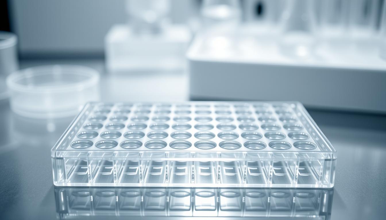
Standard Measurements for 96 Well Plates
Most culture vessels follow strict dimensional guidelines. A single well in these plates typically offers 0.32 cm², while a T-25 flask provides 25 cm². Researchers calculate seeding density per cm² to ensure uniform growth.
Medium volume ratios also matter. A well with 0.1–0.2 mL of medium maintains optimal oxygen tension. Larger formats, like 100mm dishes, use ~10 mL but require adjusted cell counts.
Comparing 96 Well Plates to Other Culture Vessels
Total usable space differs significantly across tools. A 96-well plate totals ~30.72 cm², whereas a T-75 flask offers 75 cm². Smaller formats save reagents but may compromise cell culture conditions for dense populations.
- Edge effects: Outer wells in plates evaporate faster, requiring humidity controls.
- Batch consistency: Reputable manufacturers standardize well dimensions.
- Conversion formulas: Adjust cell counts when switching between flasks and plates.
Optimizing 96 Well Surface Area for Cell Culture
Effective cell culture starts with precise adjustments to your experimental setup. Properly configuring microplates ensures uniform growth and reliable data. This section outlines actionable strategies to maximize efficiency.
Best Practices for Seeding Density and Confluency
Calculating the right number of cells per well prevents overcrowding or sparse cultures. For standard plates, aim for 0.01×10⁶ cells per well (0.32 cm²). Confluency typically peaks at 0.04×10⁶ cells.
Monitor confluency daily using microscopy or automated systems. Below are key thresholds:
| Confluency Level | Cell Count (x10⁶/well) | Recommended Action |
|---|---|---|
| 30–50% | 0.01–0.02 | Refresh medium |
| 70–90% | 0.03–0.04 | Passage cells |
| >95% | >0.04 | Risk of necrosis |
Use 0.05–0.1 mL trypsin for gentle detachment. Centrifuge at 200–300 xg for 5 minutes to pellet cells.
Balancing Volume and Surface Area for Optimal Growth
Maintain 0.1–0.2 mL medium per well to stabilize oxygen levels. Smaller volumes risk evaporation, especially in edge wells. Solutions include:
- Humidity trays to minimize evaporation.
- Frequent checks for meniscus distortion.
- Adjusted feeding schedules for high-metabolism lines.
For quality control, test plate uniformity with control wells. Address edge effects by rotating plates during incubation.
Key Factors Influencing 96 Well Plate Performance
The reliability of biological assays often depends on subtle yet critical plate characteristics. Beyond basic dimensions, material composition and engineering details determine experimental consistency. Researchers must evaluate these specifications to match plate properties with assay requirements.
Material and Surface Treatment Considerations
Polystyrene remains the standard material for most applications due to its optical clarity and cell adhesion properties. Modified versions with tissue-culture treatment enhance attachment for sensitive cell lines. Alternative materials like polypropylene offer chemical resistance for specialized research.
- Hydrophilic coatings improve cell spreading for adherent cultures
- Poly-D-lysine treatments benefit neuronal studies
- Low-protein-binding surfaces reduce background noise in ELISA
Impact of Well Shape and Design on Assay Results
Well shape dictates fluid dynamics and cell distribution patterns. Flat-bottom designs optimize microscopy, while U-bottom configurations promote uniform cell settling. Key geometric considerations include:
Curvature radius affects meniscus formation in liquid handling. Square wells maximize usable space but may create imaging artifacts. Deep-well designs accommodate larger volumes without compromising gas exchange.
Sterilization and Contamination Prevention
Gamma irradiation achieves reliable sterilization without leaving chemical residues. Validated doses (typically 25-50 kGy) maintain surface treatment integrity while eliminating microbial risks. Complementary aseptic techniques include:
– UV decontamination for work surfaces
– Positive pressure laminar flow hoods
– Sealed plate storage with desiccants
Regular contamination checks should include visual inspection and negative controls. Proper handling maintains plate performance throughout multi-day experiments.
Conclusion
Successful biological assays rely on precise plate configurations and consistent conditions. The right setup ensures accurate data and reproducible results in cell culture experiments.
Manufacturer consistency plays a vital role. Standardized well dimensions and treatments reduce variability across studies. Researchers should verify plate specifications before use.
Adapt protocols when scaling operations. Different cell types need tailored approaches for optimal growth. Continuous monitoring prevents issues like evaporation or contamination.
For deeper insights into uniform culture setups, explore this comprehensive guide on 2D model analysis. Proper planning and quality checks lead to reliable outcomes in research.
FAQ
What is the importance of surface area in cell culture plates?
Surface area directly impacts cell adhesion, growth, and nutrient exchange. Proper sizing ensures optimal confluency and reliable assay results.
How does a 96-well plate compare to other culture vessels?
These plates offer high-throughput capabilities with standardized dimensions, making them ideal for assays requiring multiple replicates in a compact format.
What materials are commonly used for manufacturing these plates?
Polystyrene is standard, often treated for cell adhesion. Specialty coatings like collagen or poly-D-lysine enhance performance for specific cell types.
How can researchers prevent contamination in multi-well plates?
Using sterile techniques, proper sealing films, and UV-treated products minimizes risks. Regular workspace decontamination is also critical.
What factors affect cell growth in high-density formats?
Seeding density, media volume, and gas exchange must be balanced. Overcrowding or insufficient nutrients can skew experimental outcomes.
Are there alternatives for specialized cell culture needs?
Yes. Low-attachment coatings or glass-bottom designs cater to sensitive applications like spheroid formation or microscopy.


Leave a Comment
Your email address will not be published. Required fields are marked *