
Successful polymerase chain reaction techniques require careful attention to multiple critical variables. Laboratory professionals must evaluate numerous factors before initiating any amplification process. These considerations directly impact the reliability and accuracy of your results.
We understand that each PCR experiment presents unique challenges. Template quality, reaction components, and thermal cycling parameters all influence amplification success. Contamination control measures and proper equipment calibration are equally essential.
Our comprehensive approach addresses the fundamental requirements for optimal molecular biology workflows. You need precise technical guidance to achieve consistent, reproducible results. We focus on delivering the expertise that enables scientific professionals to master these complex laboratory procedures with confidence and accuracy.
Key Takeaways
- Template quality and purity significantly affect amplification efficiency and specificity
- Proper primer design and concentration ratios are crucial for successful reactions
- Thermal cycling parameters must be optimized for each specific application
- Contamination prevention protocols are essential for reliable results
- Reaction component ratios require precise measurement and quality control
- Equipment calibration and maintenance directly impact experimental outcomes
Understanding the Basics of PCR
The foundation of successful PCR implementation lies in comprehending the intricate biochemical processes that govern nucleic acid detection. We provide you with essential knowledge about molecular amplification techniques that form the backbone of modern laboratory diagnostics. This understanding enables you to make informed decisions about equipment selection and protocol optimization.
PCR technology has revolutionized molecular biology by offering unprecedented precision in targeting specific genetic sequences. The technique operates through carefully controlled thermal cycling that denatures double-stranded DNA, allows primer binding, and facilitates enzymatic extension. You can achieve millions of copies from a single DNA molecule through this systematic approach.
What is Polymerase Chain Reaction?
Polymerase chain reaction represents a powerful molecular technique that amplifies specific DNA fragments through repeated thermal cycles. The process targets predetermined sequences within your sample and creates exponential increases in copy number. Each cycle consists of three distinct phases: denaturation at high temperatures, primer annealing at moderate temperatures, and extension at optimal polymerase activity temperatures.
The reaction relies on thermostable DNA polymerase enzymes that maintain functionality despite exposure to extreme temperatures. Taq polymerase remains the gold standard due to its exceptional heat resistance and reliable performance. This enzyme enables continuous cycling without requiring fresh additions between temperature changes.
Modern DNA amplification methods achieve sensitivity levels that detect single molecules in complex biological samples. The exponential nature of amplification means that 30-40 cycles can generate over one billion copies from a single template. This remarkable amplification power makes PCR indispensable for clinical diagnostics and research applications.
Components of a PCR Reaction
Every successful PCR reaction requires five essential components working in precise coordination. The template DNA provides the target sequence for amplification, while forward and reverse primers define the boundaries of the amplified region. These oligonucleotide primers must exhibit specific melting temperatures and avoid secondary structure formation.
DNA polymerase serves as the catalytic engine that synthesizes new DNA strands during the extension phase. The enzyme requires magnesium ions as cofactors and utilizes deoxynucleoside triphosphates (dNTPs) as building blocks. Buffer systems maintain optimal pH conditions and provide necessary salt concentrations for enzyme stability.
Reaction optimization involves balancing component concentrations to achieve maximum efficiency. Primer concentrations typically range from 0.1 to 1.0 μM, while dNTP levels remain around 200 μM each. Magnesium chloride concentrations require careful titration between 1.5 and 4.0 mM for optimal polymerase activity.
Types of PCR Techniques
Traditional endpoint PCR provides qualitative results through gel electrophoresis analysis after amplification completion. This approach works well for presence/absence determinations and basic molecular cloning applications. The technique offers simplicity and cost-effectiveness for routine laboratory procedures.
Real-time PCR enables quantitative analysis through fluorescent detection during amplification cycles. This advanced nucleic acid detection method provides precise quantification and reduces contamination risks. You can monitor amplification progress in real-time and obtain results within hours rather than days.
Specialized PCR variants address specific analytical challenges in different applications. Nested PCR increases sensitivity through sequential amplification rounds, while multiplex PCR allows simultaneous detection of multiple targets. Digital PCR provides absolute quantification without standard curves, offering superior precision for critical measurements.
Selecting the Right Primers
Primer selection represents one of the most critical decisions you will make when setting up PCR reactions in your molecular biology laboratory. The success of your amplification depends entirely on how well your primers bind to the target DNA sequences. We recommend following established guidelines to ensure optimal performance and reliable results.
Your primers must meet specific technical requirements to function effectively. These synthetic DNA oligonucleotides should be approximately 15-30 bases in length. This range provides sufficient specificity while maintaining efficient binding properties.
The melting temperature (Tm) of your primers should fall between 55-70°C. Both forward and reverse primers must have Tm values within 5°C of each other. This temperature matching prevents uneven amplification and ensures consistent results across your PCR cycles.
Importance of Primer Design
PCR primer design directly impacts the specificity and efficiency of your amplification reactions. Poor primer design leads to nonspecific binding, primer-dimer formation, and failed experiments. You must consider several factors when designing primers for your target sequences.
GC content represents a crucial parameter in primer design. Your primers should contain 40-60% GC content with uniform distribution throughout the sequence. This balanced composition prevents secondary structure formation and ensures stable binding to your template DNA.
Avoiding primer-dimer formation requires careful sequence analysis. These unwanted interactions occur when primers bind to each other instead of your target DNA. We recommend checking for complementarity between primer pairs during the design process.
Secondary structures within individual primers can reduce amplification efficiency. Hairpin loops and internal folding prevent proper binding to template DNA. Your primer design should minimize these structural complications.
Common Tools for Primer Design
Several bioinformatics tools streamline the PCR primer design process for laboratory applications. These software solutions analyze your target sequences and generate optimal primer pairs automatically. We recommend using multiple tools to validate your primer designs.
Primer-BLAST from NCBI provides comprehensive primer design with specificity checking. This free tool analyzes your target sequence and suggests primer pairs while checking for off-target binding sites. The interface allows you to adjust parameters based on your specific requirements.
Primer3 offers advanced primer design capabilities with extensive customization options. You can specify product size ranges, primer length preferences, and GC content requirements. This tool integrates well with other molecular biology software packages.
Commercial software packages provide additional features for complex primer design projects. These tools often include batch processing capabilities and integration with laboratory information management systems.
| Design Parameter | Recommended Range | Impact on PCR | Common Issues |
|---|---|---|---|
| Primer Length | 15-30 bases | Specificity and binding | Too short: nonspecific binding |
| Melting Temperature | 55-70°C | Annealing efficiency | Mismatch: uneven amplification |
| GC Content | 40-60% | Primer stability | Extreme values: poor binding |
| Product Size | 100-1000 bp | Amplification success | Too long: incomplete extension |
Quality primer design software also checks for potential secondary structures and primer-dimer interactions. These features help you avoid common pitfalls that compromise PCR performance in your laboratory workflows.
Optimizing PCR Conditions
PCR optimization involves precise control of thermal cycling parameters and reagent concentrations to maximize amplification efficiency. We provide comprehensive strategies to help you achieve consistent, high-quality results through systematic condition optimization. Understanding the relationship between temperature profiles, chemical components, and polymerase selection forms the foundation of successful PCR troubleshooting.
Laboratory professionals face common challenges including poor yield, nonspecific amplification, and primer-dimer formation. These issues often stem from suboptimal reaction conditions rather than experimental design flaws. By following proven optimization protocols, you can systematically address these problems and improve your amplification success rates.
Temperature and Time Profiles
The thermal cycling protocol represents the most critical aspect of PCR optimization. Denaturation begins at 95°C to disrupt hydrogen bonds between DNA strands, ensuring complete template separation. This high-temperature step typically requires 15-30 seconds for effective strand separation.
Annealing temperatures range from 55°C to 72°C, depending on primer melting temperatures and target specificity requirements. We recommend starting 5°C below the calculated primer melting temperature and adjusting based on results. Proper annealing temperature selection prevents nonspecific binding while ensuring efficient primer hybridization.
Extension occurs at 75°C to 80°C, with timing based on target length and polymerase processivity. Standard Taq polymerase synthesizes approximately 1000 base pairs per minute at optimal temperature. Longer targets require proportionally extended extension times to ensure complete synthesis.
MgCl2 Concentration
Magnesium concentration directly affects polymerase activity and primer binding specificity. Optimal concentrations typically range from 1-4 mM, with most reactions performing best between 1.5-2.5 mM. PCR troubleshooting often begins with magnesium optimization due to its significant impact on reaction success.
Low magnesium concentrations below 1 mM result in reduced or absent PCR products. Insufficient Mg2+ levels prevent proper polymerase function and reduce primer binding efficiency. Conversely, excessive magnesium above 4 mM promotes nonspecific amplification and primer-dimer formation.
We recommend titrating magnesium in 0.5 mM increments to identify optimal concentrations for your specific primers and templates. This systematic approach ensures you achieve maximum specificity while maintaining adequate amplification efficiency. Document successful magnesium concentrations for future experiments using similar primer sets.
Effect of DNA Polymerase Type
Polymerase selection significantly influences PCR performance and product quality. Standard Taq polymerase offers cost-effective amplification for routine applications but lacks 3′-5′ exonuclease activity. This limitation results in higher error rates compared to high-fidelity enzymes.
High-fidelity polymerases incorporate proofreading activity, reducing error rates by 10-50 fold compared to standard Taq. These enzymes prove essential for cloning applications where sequence accuracy is critical. However, they typically require longer extension times and may show reduced efficiency with certain templates.
Hot-start polymerases remain inactive at room temperature, preventing nonspecific amplification during reaction setup. This feature improves specificity and reduces primer-dimer formation in challenging reactions. Consider hot-start variants when working with low-abundance targets or complex templates.
| Parameter | Standard Range | Optimization Strategy | Common Issues |
|---|---|---|---|
| Denaturation Temperature | 94-96°C | Use 95°C for most applications | Incomplete denaturation at low temps |
| Annealing Temperature | 55-72°C | Start 5°C below primer Tm | Nonspecific binding, poor efficiency |
| Extension Temperature | 72-78°C | Use polymerase-specific optimum | Incomplete synthesis, reduced yield |
| MgCl2 Concentration | 1.0-4.0 mM | Titrate in 0.5 mM increments | No product or nonspecific bands |
Successful optimization requires systematic testing of individual parameters while maintaining consistent experimental conditions. Start with recommended ranges and adjust based on your specific requirements. Document successful conditions for reproducible results across multiple experiments.
The thermal cycling protocol optimization process typically requires 3-5 test runs to achieve optimal conditions. This investment in initial optimization saves time and resources in subsequent experiments. Remember that different primer pairs may require unique optimization even within the same laboratory.
Sample Quality and Preparation
The integrity of your DNA samples directly determines the reliability of quantitative PCR results. We understand that proper sample preparation serves as the cornerstone of successful laboratory experiments. High-quality templates ensure accurate amplification and minimize the risk of false results that can compromise your research outcomes.
Sample preparation involves multiple critical steps that affect nucleic acid detection accuracy. Each stage requires careful attention to detail and adherence to established protocols. The quality of your starting material influences every subsequent step in the PCR process.
How to Isolate DNA
DNA isolation requires systematic approaches that preserve template integrity while removing cellular debris. We recommend starting with fresh samples whenever possible to maintain optimal nucleic acid quality. The isolation process typically involves cell lysis, protein removal, and DNA purification steps.
Several purification methods effectively prepare DNA for quantitative PCR applications:
- Ethanol precipitation – Concentrates DNA while removing salts and small molecules
- Dialysis – Eliminates low molecular weight contaminants through membrane separation
- Chloroform extraction – Removes proteins and lipids from aqueous DNA solutions
- Chromatography – Provides high-purity DNA through column-based separation
Each method offers specific advantages depending on your sample type and downstream applications. The choice of purification technique should align with your experimental requirements and available laboratory resources.
Impact of Contaminants on PCR
Common inhibitors significantly affect PCR performance and nucleic acid detection sensitivity. We have identified several substances that interfere with polymerase activity and template amplification. Understanding these contaminants helps you implement appropriate removal strategies.
Major PCR inhibitors include:
- Proteinase K – Residual enzyme activity can degrade polymerase
- Phenol – Organic solvent that denatures proteins and enzymes
- EDTA – Chelates essential magnesium ions required for polymerase function
- Ionic detergents – Disrupt enzyme structure and membrane integrity
- Heparin – Anticoagulant that binds to polymerase and blocks activity
Additional inhibitors such as spermidine and hemoglobin can also compromise reaction efficiency. These substances often originate from biological samples or carry over from extraction procedures. Proper purification removes most contaminants and ensures reliable quantitative PCR results.
Storage Conditions for DNA Samples
Proper storage maintains DNA integrity and prevents degradation that affects nucleic acid detection accuracy. We recommend specific conditions based on storage duration and sample requirements. Temperature control represents the most critical factor in preserving template quality.
Short-term storage at 4°C works well for samples used within one week. Long-term preservation requires freezing at -20°C or -80°C depending on stability requirements. Buffer systems containing EDTA help prevent nuclease activity during storage.
Avoid repeated freeze-thaw cycles that fragment DNA and reduce template quality. Aliquot samples into small volumes to minimize temperature fluctuations. Proper labeling and documentation ensure sample traceability and prevent mix-ups that compromise experimental integrity.
Setting Up a PCR Reaction
Effective PCR reaction setup combines technical precision with comprehensive contamination prevention strategies in molecular biology laboratory environments. We provide essential protocols that ensure accurate results while maintaining the highest standards of quality control. Your success depends on systematic preparation, proper equipment selection, and strict adherence to established procedures.
The foundation of reliable amplification begins with understanding the critical factors that influence reaction success. Temperature control, reagent quality, and workspace organization directly impact your experimental outcomes. Each component requires careful consideration to achieve consistent, reproducible results.
Pipetting Techniques for Accuracy
Precise pipetting forms the cornerstone of successful PCR experiment execution. Positive displacement pipettes provide superior accuracy when handling viscous solutions and small volumes. These instruments eliminate air displacement errors that commonly affect standard pipettes during critical measurements.
We recommend using barrier tips for all PCR-related pipetting activities. These specialized tips prevent aerosol contamination and cross-contamination between samples. The barrier membrane blocks liquid and vapor from entering the pipette shaft, maintaining reaction integrity throughout your molecular biology laboratory workflow.
Temperature equilibration of reagents improves pipetting accuracy significantly. Allow all components to reach room temperature before use, except for heat-sensitive enzymes. Pre-wet pipette tips with the solution being transferred to ensure accurate volume delivery, particularly with small volumes below 10 microliters.
Required Equipment and Reagents
Essential equipment for PCR experiment setup includes dedicated pipettes, thermal cyclers, and specialized consumables. Your molecular biology laboratory should maintain separate sets of equipment for pre-PCR and post-PCR activities to prevent contamination. This segregation protects reaction integrity and ensures reliable results.
Quality reagents form the foundation of successful amplification reactions. High-grade DNA polymerases with proofreading activity reduce error rates and improve specificity. Buffer systems must maintain optimal pH and ionic strength throughout thermal cycling conditions.
| Equipment Category | Essential Items | Quality Requirements | Contamination Risk |
|---|---|---|---|
| Pipetting Systems | Positive displacement pipettes, barrier tips, multichannel pipettes | Calibrated annually, accuracy ±2% | High |
| Thermal Equipment | Thermal cycler, heating blocks, ice buckets | Temperature uniformity ±0.5°C | Medium |
| Reagent Storage | Freezers, refrigerators, desiccant chambers | Temperature stability ±1°C | Low |
| Consumables | PCR tubes, plates, sealing films | DNase/RNase free, certified | High |
Master mix preparation requires careful attention to component ratios and mixing procedures. Prepare master mixes on ice to maintain enzyme stability. Vortex gently and centrifuge briefly to ensure homogeneous distribution without introducing air bubbles that affect reaction volumes.
Best Practices for Contamination Control
Contamination prevention represents the most critical aspect of PCR experiment success in any molecular biology laboratory. Physical separation of pre-PCR and post-PCR areas prevents amplicon contamination that can generate false positive results. Dedicated equipment, reagents, and personnel flow patterns maintain this separation effectively.
Personal protective equipment requirements include face masks, gloves, and hair covers worn consistently throughout the procedure. Change gloves between samples and after handling any potentially contaminated surfaces. Disposable lab coats provide additional protection and should be changed when moving between different laboratory areas.
UV irradiation of work surfaces and equipment helps eliminate residual DNA contamination. Expose surfaces to UV light for 15-30 minutes before beginning work. However, avoid UV exposure of reagents and samples, as this can cause DNA damage and reduce PCR experiment efficiency.
Workflow organization minimizes contamination risks through logical sequence planning. Process negative controls first, followed by samples in order of expected template concentration. Positive controls should be handled last to prevent high-concentration template from contaminating other reactions.
Environmental controls maintain optimal conditions for accurate results. Laminar flow hoods provide clean air environments for sensitive procedures. Regular HEPA filter maintenance and airflow verification ensure consistent performance. Temperature and humidity monitoring prevents condensation that can affect reagent stability and pipetting accuracy.
We emphasize the importance of establishing standard operating procedures for your molecular biology laboratory. Document all protocols, equipment maintenance schedules, and quality control measures. Regular training updates ensure all personnel maintain current best practices for contamination prevention and accurate PCR experiment execution.
PCR Cycle Parameters
Thermal cycling protocol optimization forms the foundation of successful DNA amplification methods in laboratory settings. We provide comprehensive guidance on mastering the three critical phases that define PCR success. You need precise control over temperature profiles, timing sequences, and cycle numbers to achieve consistent amplification results.
The thermal cycling process involves repeated temperature changes that enable DNA replication. Each cycle multiplies your target DNA sequence exponentially. Understanding these parameters helps you troubleshoot issues and optimize your experimental outcomes.
Understanding Denaturation, Annealing, and Extension
The denaturation phase occurs at 94-98°C for 15-30 seconds. This high temperature separates double-stranded DNA into single strands. You must ensure complete denaturation to prevent incomplete amplification and poor product yield.
Annealing happens at 50-65°C for 20-40 seconds. Your primers bind to their complementary sequences during this phase. The annealing temperature depends on primer length, GC content, and sequence specificity.
Extension takes place at 72°C for 30 seconds to 2 minutes. DNA polymerase synthesizes new DNA strands during this phase. Extension time varies based on your target sequence length and polymerase efficiency.
- Short targets (under 500 bp): 30-45 seconds extension
- Medium targets (500-2000 bp): 1-2 minutes extension
- Long targets (over 2000 bp): 2+ minutes extension
Cycle Number Considerations
Most thermal cycling protocols require 25-40 cycles for optimal results. Amplification efficiency declines after 30-40 cycles due to several factors. Reagent depletion becomes significant at higher cycle numbers.
You should consider these limiting factors in your DNA amplification methods:
- Enzyme activity reduction from repeated heating
- Accumulation of inhibitory byproducts
- Competition between products and templates
- Primer and nucleotide depletion
We recommend starting with 30 cycles for most applications. Increase cycle numbers gradually if amplification appears insufficient. Excessive cycling can lead to non-specific products and reduced specificity.
Troubleshooting PCR Cycle Issues
Poor amplification often results from incorrect thermal cycling protocol parameters. You can identify problems by analyzing your temperature profiles and timing sequences. We help you systematically address common cycling issues.
Incomplete denaturation causes weak bands and inconsistent results. Increase denaturation temperature by 2-4°C or extend denaturation time. Check your thermal cycler calibration regularly.
Poor primer annealing leads to low specificity and multiple bands. Optimize annealing temperature using gradient PCR. Calculate melting temperatures accurately for your primer pairs.
Insufficient extension time results in incomplete products and reduced yield. Extend your extension phase by 30-60 seconds for longer targets. Consider using high-fidelity polymerases for improved efficiency.
| Problem | Cause | Solution | Expected Result |
|---|---|---|---|
| No amplification | Denaturation too low | Increase to 95-98°C | Clear product bands |
| Multiple bands | Annealing too low | Raise annealing temperature | Single specific band |
| Weak products | Too few cycles | Add 5-10 more cycles | Stronger band intensity |
| Smeared bands | Extension too short | Extend by 30-60 seconds | Sharp, defined bands |
Analyzing PCR Results
Proper analysis of PCR results requires systematic evaluation of amplification products through established visualization techniques. We provide comprehensive protocols that ensure accurate interpretation of your experimental outcomes. The success of any PCR experiment depends on your ability to correctly assess the quality and specificity of amplified DNA products.
After completing the amplification process, you must verify that your PCR reaction ingredients produced the expected results. This verification process involves multiple analytical steps that confirm both the presence and purity of your target DNA sequences.
Techniques for Gel Electrophoresis
Agarose gel electrophoresis serves as the primary method for visualizing PCR products. You prepare the gel by dissolving agarose powder in buffer solution, typically at concentrations between 0.8% and 2.0% depending on your expected fragment sizes.
The gel electrophoresis analysis process involves several critical steps:
- Sample preparation: Mix your PCR products with loading dye containing tracking dyes and density agents
- Gel loading: Carefully pipette samples into wells alongside molecular weight markers
- Electrophoresis conditions: Run the gel at appropriate voltage (typically 80-120V) for optimal separation
- Staining and visualization: Use ethidium bromide or safer alternatives like SYBR Safe for DNA detection
We recommend using ultraviolet transilluminators for visualizing stained DNA bands. However, you should always follow proper safety protocols when working with UV light and DNA intercalating dyes.
Understanding Band Sizes and Purity
Accurate interpretation of your gel electrophoresis analysis results requires understanding molecular weight markers and band migration patterns. DNA fragments migrate through the gel matrix based on their size, with smaller fragments traveling farther than larger ones.
You can determine fragment sizes by comparing your PCR products to standard molecular weight ladders. Common ladder sizes include 100bp, 1kb, and 50bp increments that provide reference points for size estimation.
Band intensity indicates the relative amount of PCR product present. Strong, sharp bands typically suggest successful amplification, while weak or smeared bands may indicate:
- Insufficient template DNA concentration
- Suboptimal primer annealing conditions
- DNA degradation during storage or handling
- Incomplete amplification cycles
Product purity assessment involves examining band sharpness and the absence of additional unwanted bands. Single, discrete bands at the expected molecular weight indicate high-purity PCR products suitable for downstream applications.
Common Issues in PCR Analysis
Effective PCR troubleshooting requires systematic identification of common analytical problems. We help you recognize and resolve issues that can compromise result interpretation and experimental reliability.
Primer-dimer formation appears as low molecular weight bands (typically 50-100bp) at the bottom of your gel. These artifacts result from primer self-annealing and can be minimized through optimized reaction conditions and hot-start polymerases.
Nonspecific amplification manifests as multiple bands of varying sizes beyond your target product. This issue often stems from:
- Excessive primer concentrations
- Low annealing temperatures
- High template DNA amounts
- Insufficient reaction specificity
Contamination artifacts present unique challenges in PCR troubleshooting. Cross-contamination between samples produces unexpected bands, while reagent contamination can generate false-positive results. Implementing proper negative controls helps identify these contamination sources.
Smeared or streaked bands indicate DNA degradation or incomplete denaturation. You can address these issues by verifying template quality, optimizing denaturation temperatures, and ensuring proper storage conditions for all reaction components.
Southern blot hybridization provides additional verification for result specificity when gel electrophoresis analysis alone proves insufficient. This technique involves transferring DNA from the gel to a membrane and probing with specific labeled sequences that confirm target identity.
We recommend maintaining detailed documentation of all analytical procedures and results. This practice supports reproducibility and facilitates effective PCR troubleshooting when unexpected results occur in future experiments.

Practical Applications of PCR
The practical applications of PCR extend far beyond basic laboratory procedures, encompassing critical roles in healthcare, scientific research, and criminal justice systems. We observe how nucleic acid detection technologies have transformed diagnostic capabilities across multiple industries. Modern laboratories depend on these versatile techniques to solve complex analytical challenges.
PCR applications demonstrate remarkable adaptability in addressing diverse scientific needs. You will find these technologies essential for pathogen identification, genetic analysis, and forensic investigations. The reliability and precision of PCR make it indispensable for professionals requiring accurate molecular detection.
Clinical Diagnostics and Pathogen Detection
Clinical laboratories utilize PCR for comprehensive pathogen detection and disease diagnosis. Quantitative PCR enables precise identification of viral, bacterial, fungal, and parasitic organisms in patient samples. Healthcare professionals rely on these methods for rapid and accurate diagnostic results.
Viral pathogen detection represents a primary application area for clinical PCR. We support laboratories in detecting human papillomavirus, HIV, herpes simplex virus, and SARS-CoV-2 through specialized protocols. These applications require stringent quality control measures to ensure diagnostic accuracy.
Additional viral targets include varicella-zoster virus, enterovirus, cytomegalovirus, and hepatitis viruses. Each pathogen requires specific primer designs and optimized reaction conditions. Clinical laboratories must maintain validated protocols for consistent performance.
Bacterial and fungal detection through nucleic acid detection provides rapid identification compared to traditional culture methods. PCR reduces diagnostic timeframes from days to hours. This speed improvement directly impacts patient care and treatment decisions.
Parasitic organism identification benefits from PCR sensitivity and specificity. Traditional microscopy methods often miss low-level infections. PCR detection enables early intervention and appropriate treatment protocols.
Research and Development Uses
Research laboratories employ PCR for diverse molecular biology applications. Gene expression analysis through quantitative PCR provides precise measurement of RNA levels. Scientists use these techniques to study cellular responses and regulatory mechanisms.
Point mutation detection represents a critical research application. PCR enables identification of single nucleotide changes in genetic sequences. This capability supports genetic disorder research and therapeutic development programs.
DNA sequencing preparation relies heavily on PCR amplification. Researchers amplify target regions before sequencing analysis. This approach ensures sufficient template material for accurate sequence determination.
In vitro mutagenesis applications utilize PCR for introducing specific genetic changes. Scientists create modified DNA sequences for functional studies. These techniques advance our understanding of gene function and protein structure.
Molecular cloning procedures incorporate PCR for vector preparation and insert amplification. Research teams use these methods to construct recombinant DNA molecules. PCR streamlines cloning workflows and improves success rates.
Forensic Science Applications
Forensic laboratories depend on PCR for DNA profiling and criminal investigations. Nucleic acid detection enables analysis of degraded or limited biological samples. These capabilities prove essential for solving complex criminal cases.
DNA profiling through PCR amplification generates genetic fingerprints from trace evidence. Forensic experts analyze short tandem repeat markers for individual identification. This technology provides reliable evidence for legal proceedings.
Paternity testing applications utilize PCR for genetic relationship determination. Laboratories compare DNA profiles between alleged parents and children. These analyses provide definitive answers for legal and personal situations.
Criminal investigation support includes analysis of biological evidence from crime scenes. PCR enables DNA recovery from challenging samples including hair, saliva, and blood traces. Forensic teams rely on these methods for case resolution.
| Application Field | Primary Use Cases | Key Benefits | Sample Types |
|---|---|---|---|
| Clinical Diagnostics | Pathogen detection, disease diagnosis, viral load monitoring | Rapid results, high sensitivity, quantitative analysis | Blood, tissue, swabs, body fluids |
| Research & Development | Gene expression, mutation detection, cloning, sequencing | Precision measurement, versatile protocols, scalable methods | Cell cultures, tissue samples, purified DNA/RNA |
| Forensic Science | DNA profiling, paternity testing, criminal investigation | Trace sample analysis, degraded DNA recovery, legal reliability | Hair, saliva, blood, touch DNA, bone fragments |
| Environmental Testing | Microbial monitoring, species identification, contamination detection | Environmental sample compatibility, field applications, rapid screening | Water, soil, air filters, plant material |
We recognize the expanding role of PCR technology across these diverse application areas. Each field presents unique challenges requiring specialized approaches and validated methodologies. Our expertise supports laboratories in implementing appropriate PCR solutions for their specific analytical needs.
The versatility of quantitative PCR continues to drive innovation in diagnostic and research applications. New developments in primer design, reaction chemistry, and detection methods enhance performance capabilities. These advances expand the potential applications for nucleic acid detection technologies.
Quality assurance remains paramount across all PCR applications. Laboratories must maintain rigorous validation protocols and quality control measures. We provide comprehensive support for establishing and maintaining these critical quality systems.
Comparison of PCR Variants
The evolution of polymerase chain reaction techniques has created multiple specialized variants, each designed for specific laboratory requirements. Modern molecular biology laboratories must evaluate these different methodologies to select the most appropriate approach for their research objectives. We provide comprehensive analysis to help you understand the distinct advantages and limitations of each PCR variant.
Each PCR methodology offers unique capabilities that address specific analytical challenges. Your choice depends on factors such as sensitivity requirements, quantification needs, budget constraints, and time considerations. Understanding these differences enables you to optimize your experimental workflows and achieve more reliable results.
Real-Time PCR vs. Traditional PCR
Real-time PCR represents a significant advancement over traditional endpoint PCR methods. This technology allows immediate detection of amplified products during the reaction through incorporation of fluorescent molecules. You can monitor amplification in real-time, providing quantitative data throughout the process.
Traditional PCR requires gel electrophoresis for result analysis after completion of all cycles. This approach involves additional time and labor for post-amplification processing. Real-time PCR eliminates these extra steps by providing immediate results through fluorescent detection systems.
The cost difference between these methods is substantial. Real-time PCR systems require specialized instrumentation and fluorescent reagents, making them more expensive than conventional PCR setups. However, the enhanced speed and quantification capabilities often justify the additional investment for high-throughput applications.
Quantification accuracy represents another key distinction. Real-time PCR provides precise quantitative measurements through cycle threshold (Ct) values. Traditional PCR offers only qualitative or semi-quantitative results through band intensity analysis on gels.
Digital PCR: Advantages and Limitations
Digital PCR provides absolute quantification without requiring standard curves or reference samples. This technology partitions your sample into thousands of individual reactions, enabling precise counting of target molecules. The superior precision makes digital PCR ideal for rare target detection and copy number variation analysis.
The main advantage of digital PCR lies in its ability to provide absolute quantification. You obtain exact molecule counts rather than relative measurements. This capability proves particularly valuable for clinical diagnostics and quality control applications where precise quantification is critical.
However, digital PCR has notable limitations. The technology requires specialized equipment that represents a significant capital investment. Sample throughput is typically lower compared to real-time PCR systems. Additionally, the dynamic range is more limited, making it less suitable for samples with very high target concentrations.
Cost per reaction is generally higher for digital PCR due to the specialized consumables required. The analysis time is also longer compared to real-time PCR methods. These factors must be weighed against the enhanced precision and absolute quantification capabilities.
Nested PCR: When to Use It
Nested PCR employs two rounds of amplification using different primer sets to achieve enhanced sensitivity and specificity. This approach proves particularly valuable when working with degraded samples or detecting low-abundance targets. The two-step amplification process significantly reduces non-specific products while increasing target sensitivity.
You should consider nested PCR when standard PCR fails to detect your target due to low template concentration. This method excels in forensic applications, ancient DNA analysis, and pathogen detection from clinical samples with low viral loads. The enhanced specificity also makes it suitable for detecting specific genetic variants.
The main disadvantage of nested PCR is increased contamination risk. The additional handling steps and tube opening create more opportunities for cross-contamination. Careful laboratory practices and dedicated workspace areas become essential when implementing nested PCR protocols.
Time requirements are significantly higher for nested PCR compared to single-round amplification. You must complete two separate PCR reactions, effectively doubling the processing time. This factor makes nested PCR less suitable for high-throughput applications or time-sensitive analyses.
| PCR Variant | Sensitivity | Quantification | Cost | Time Required |
|---|---|---|---|---|
| Traditional PCR | Moderate | Qualitative | Low | 3-4 hours |
| Real-Time PCR | High | Quantitative | Moderate | 1-2 hours |
| Digital PCR | Very High | Absolute | High | 2-3 hours |
| Nested PCR | Very High | Qualitative | Low | 6-8 hours |
Selecting the appropriate PCR variant requires careful consideration of your specific analytical requirements. We recommend evaluating sensitivity needs, quantification requirements, budget constraints, and time limitations before making your decision. For comprehensive guidance on different types of PCR techniques and their, consider your laboratory’s long-term objectives and available resources.
Each polymerase chain reaction technique serves distinct purposes in modern molecular biology laboratory settings. Your optimal choice depends on balancing performance requirements with practical considerations such as cost, time, and available expertise.
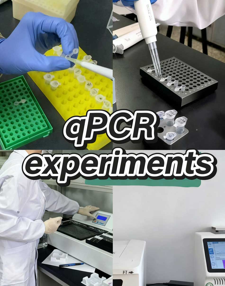
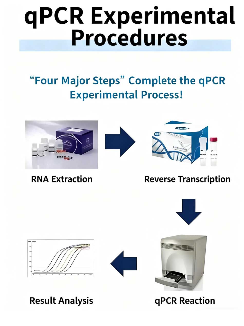
gPCR Experimental Procedures
Case Studies in PCR Implementation
Evidence-based examples illustrate how PCR technology enables breakthrough solutions in healthcare, research, and environmental monitoring. We present comprehensive case studies that demonstrate successful implementation of DNA amplification methods across diverse scientific applications. These real-world scenarios showcase the versatility and impact of nucleic acid detection technologies in addressing complex analytical challenges.
Laboratory professionals worldwide have witnessed transformative results through strategic PCR implementation. The following case studies provide concrete evidence of how this technology revolutionizes diagnostic capabilities and research outcomes.
Successful Uses in Infectious Disease Outbreaks
The COVID-19 pandemic exemplifies PCR’s critical role in global health management. RT-PCR served as the primary diagnostic method due to its exceptional sensitivity, specificity, and rapid turnaround time. Healthcare systems relied on this nucleic acid detection approach to identify infected individuals within hours rather than days.
Clinical laboratories successfully implemented PCR protocols to detect multiple bacterial pathogens simultaneously. DNA amplification methods enabled precise identification of Mycobacterium species, Leptospira genospecies, and Chlamydia species in patient samples. This rapid diagnostic capability facilitated early treatment decisions and improved patient outcomes.
Public health agencies utilized PCR technology for outbreak investigation and control. The detection of Legionella pneumophila, Listeria monocytogenes, and Neisseria meningitidis through targeted amplification supported source tracing efforts. Laboratory teams achieved turnaround times of 2-4 hours, enabling swift implementation of containment measures.
Contact tracing programs benefited significantly from PCR’s analytical precision. Healthcare workers could identify asymptomatic carriers and implement quarantine protocols effectively. The technology’s ability to detect low viral loads proved essential for preventing transmission chains.
Advances in Genetic Research through PCR
Rare disease diagnosis has been revolutionized through innovative DNA amplification methods. Research laboratories successfully identified genetic mutations in patients with previously undiagnosed conditions. Case studies demonstrate how targeted PCR approaches reduced diagnostic timelines from months to weeks.
Pharmacogenomics applications showcase PCR’s impact on personalized medicine development. Clinical researchers implemented nucleic acid detection protocols to identify drug metabolism variants in patient populations. These findings enabled healthcare providers to optimize medication dosing and reduce adverse reactions.
Cancer research laboratories achieved breakthrough results through PCR-based genetic profiling. Scientists successfully identified tumor-specific mutations that guided targeted therapy selection. The technology’s precision enabled detection of circulating tumor DNA in blood samples, facilitating non-invasive monitoring approaches.
Prenatal genetic screening programs expanded their capabilities through advanced PCR implementations. Laboratory teams developed protocols for detecting chromosomal abnormalities with unprecedented accuracy. These applications reduced the need for invasive diagnostic procedures while maintaining diagnostic reliability.
Environmental Applications of PCR
Water quality monitoring programs demonstrate PCR’s environmental impact through pathogen detection initiatives. Municipal laboratories successfully implemented DNA amplification methods to identify waterborne pathogens in drinking water supplies. These applications enabled rapid response to contamination events and protected public health.
Food safety testing laboratories revolutionized their analytical capabilities through PCR implementation. Quality control teams achieved same-day detection of foodborne pathogens including Salmonella, E. coli, and Campylobacter species. The technology’s sensitivity enabled detection of contamination at levels previously undetectable through traditional methods.
Biodiversity assessment projects utilized nucleic acid detection approaches for species identification in environmental samples. Research teams successfully analyzed soil and water samples to catalog microbial communities. These studies provided critical data for ecosystem health monitoring and conservation efforts.
Agricultural laboratories implemented PCR protocols for plant pathogen detection and crop protection. Scientists developed rapid screening methods for viral, bacterial, and fungal diseases affecting major food crops. The technology’s specificity enabled targeted treatment approaches that reduced pesticide usage while maintaining crop yields.
Environmental forensics applications showcase PCR’s versatility in contamination source identification. Regulatory agencies successfully traced pollution sources through genetic fingerprinting of microbial communities. These investigations supported enforcement actions and remediation planning efforts across diverse environmental settings.
Future Trends in PCR Technology
The landscape of molecular biology continues to evolve as breakthrough technologies reshape how we approach PCR experiment design and implementation. These advances promise to streamline laboratory workflows while enhancing analytical capabilities across research and clinical settings.
Innovations in PCR Methodologies
Automated sample-to-result systems represent the next generation of PCR technology. Leading instrument manufacturers are developing completely self-contained platforms that eliminate the need for separate preparation and analysis rooms. These integrated systems reduce contamination risks while improving workflow efficiency.
Miniaturized PCR platforms offer rapid thermal cycling with reduced reagent consumption. Digital PCR technologies provide absolute quantification without standard curves, enabling more precise measurements for critical applications.
Impact of AI on PCR Processes
Artificial intelligence algorithms now optimize PCR conditions automatically by analyzing amplification curves and adjusting parameters in real-time. Machine learning models predict optimal primer designs and identify potential issues before they affect results.
Smart data analysis tools interpret gel electrophoresis analysis results with enhanced accuracy, reducing human error in band identification and size determination.
The Role of PCR in Precision Medicine
Personalized treatment approaches increasingly rely on PCR-based companion diagnostics. These tests guide therapeutic selection by identifying specific genetic markers that predict drug response.
Real-time monitoring of treatment efficacy through circulating biomarkers enables dynamic therapy adjustments. Individual tube analysis during real-time PCR allows for patient-specific optimization of diagnostic protocols, supporting the shift toward truly personalized healthcare solutions.
References and further readings:
1.Soltani S, Erami M, Ahmadikia K, Aboutalebian S, Rezaie S, Badali H. Molecular assays versus mycological methods for diagnosis of rhino-orbital mucormycosis: analysis of 120 clinical specimens from COVID-19 patients. Mycopathologia. 2025.
https://link.springer.com/article/10.1007/s11046-025-00937-72.Devonshire AS, Morata J, Jubin C, Abreu Pereira RP, Bolognini D, Seitz H. Interlaboratory evaluation of high molecular weight DNA extraction methods for long-read sequencing and structural variant analysis. BMC Genomics. 2025. doi:10.1186/s12864-025-11792-7
https://link.springer.com/article/10.1186/s12864-025-11792-73.Zlender T, Brezočnik L, Podgorelec V, Rupnik M. MicrobiomePrime: A primer pair selection tool for microbial source tracking validated on a comprehensive collection of animal gut and fecal waste microbiomes. bioRxiv. 2025.
https://www.biorxiv.org/content/10.1101/2025.07.08.663663v14.Li J, Zhang K, Han Y, Peng R, Li L, Li J. A novel method for the preparation of reproducible, stable, and non-infectious quality control materials for Chlamydia trachomatis nucleic acid detection. Microbiology Spectrum. 2024;12(4).
https://journals.asm.org/doi/10.1128/spectrum.00837-24
FAQ
What are the most critical factors to consider before starting a PCR experiment?
We recommend evaluating four fundamental parameters: template quality (ensuring DNA integrity and purity), reaction components (proper concentrations of primers, polymerase, and nucleotides), thermal cycling conditions (optimized temperature profiles and timing), and contamination control measures (sterile techniques and workspace separation). These factors directly influence amplification success and experimental reproducibility in molecular biology laboratory settings.
How does the polymerase chain reaction work at the molecular level?
PCR operates through a three-phase amplification process: denaturation (separating DNA strands at high temperature), annealing (primers binding to target sequences at lower temperature), and extension (DNA polymerase synthesizing new strands). This cycle repeats 25-40 times, exponentially amplifying the target DNA sequence. The process requires thermostable enzymes, template DNA, primers, and nucleotides to achieve successful DNA amplification methods.
What specifications should I follow for optimal PCR primer design?
We recommend primers of 18-25 nucleotides in length with melting temperatures between 55-65°C and GC content of 40-60%. Avoid primer-dimer formation, secondary structures, and nonspecific binding sites. Use bioinformatics tools like Primer3 or NCBI Primer-BLAST for design optimization. Proper primer design ensures amplification specificity and consistent results across diverse experimental conditions.
How do I optimize thermal cycling protocol parameters for maximum PCR efficiency?
Start with denaturation at 94-98°C for 15-30 seconds, annealing 2-5°C below primer Tm for 15-60 seconds, and extension at 72°C for 1 minute per kb of product. Optimize magnesium concentrations (typically 1.5-2.5 mM) and select appropriate DNA polymerase based on your application. Systematically adjust parameters while monitoring amplification efficiency and specificity through gel electrophoresis analysis.
What sample preparation steps are essential for reliable quantitative PCR results?
Ensure high-quality DNA isolation using appropriate extraction methods, assess template concentration and purity (A260/A280 ratio 1.8-2.0), and remove PCR inhibitors through purification techniques. Store DNA samples at -20°C for short-term or -80°C for long-term storage. Maintain proper buffer systems and avoid freeze-thaw cycles that can compromise template integrity and affect nucleic acid detection accuracy.
What are the best practices for contamination control in PCR reaction setup?
Establish separate pre-PCR and post-PCR areas, use dedicated pipettes and equipment for each zone, and implement proper master mix preparation protocols. Always include negative controls, use filter tips, and maintain sterile workspace conditions. Follow systematic template addition procedures and organize reagents to minimize cross-contamination risks throughout the molecular biology laboratory workflow.
How do I troubleshoot common PCR cycle issues like poor amplification?
Address incomplete denaturation by increasing temperature or time, resolve poor primer annealing by optimizing annealing temperature, and ensure sufficient extension time for your target length. Check magnesium concentrations, verify primer quality, and assess template integrity. Consider PCR troubleshooting approaches including gradient PCR for temperature optimization and enzyme concentration adjustments for improved performance.
What techniques should I use for proper gel electrophoresis analysis of PCR products?
Use 1-2% agarose gels for most applications, run at appropriate voltage (5-10 V/cm), and include molecular weight markers for band size determination. Visualize DNA using ethidium bromide or safer alternatives like SYBR Safe. Identify primer-dimers (typically
What are the main applications of PCR in clinical diagnostics?
PCR enables pathogen identification (bacterial, viral, fungal), genetic disorder screening, and infectious disease monitoring in clinical settings. Applications include COVID-19 detection through RT-PCR, bacterial resistance gene identification, and hereditary disease diagnosis. The technology supports rapid diagnostic workflows, enabling timely treatment decisions and public health responses in healthcare environments.
How does real-time PCR compare to traditional endpoint PCR methods?
Real-time PCR provides quantitative results, faster turnaround times, and reduced contamination risk through closed-tube systems. It offers superior precision for quantitative PCR applications and enables real-time monitoring of amplification. Traditional PCR requires gel electrophoresis for analysis but costs less initially. Choose based on your need for quantification, speed, and budget considerations in your molecular biology laboratory.
Can you provide examples of successful PCR implementation in disease outbreaks?
PCR technology proved critical during the COVID-19 pandemic for rapid SARS-CoV-2 detection, enabling widespread testing and contact tracing. Previous applications include MERS-CoV identification, H1N1 influenza detection, and bacterial pathogen identification in foodborne illness outbreaks. These implementations demonstrate PCR’s versatility in emergency response scenarios and public health protection through accurate nucleic acid detection.
What future innovations are transforming PCR technology and laboratory workflows?
Emerging developments include automated sample-to-result systems, miniaturized PCR platforms, and AI-enhanced optimization algorithms for improved efficiency. Machine learning applications enable predictive modeling for primer design and result interpretation. Advanced detection technologies are reducing analysis time while increasing sensitivity. These innovations support precision medicine applications and personalized treatment approaches in modern healthcare settings.
Leo Bios
Hello, I’m Leo Bios. As an assistant lecturer, I teach cellular and
molecular biology to undergraduates at a regional US Midwest university. I started as a research tech in
a biotech startup over a decade ago, working on molecular diagnostic tools. This practical experience
fuels my teaching and writing, keeping me engaged in biology’s evolution.


Leave a Comment
Your email address will not be published. Required fields are marked *