


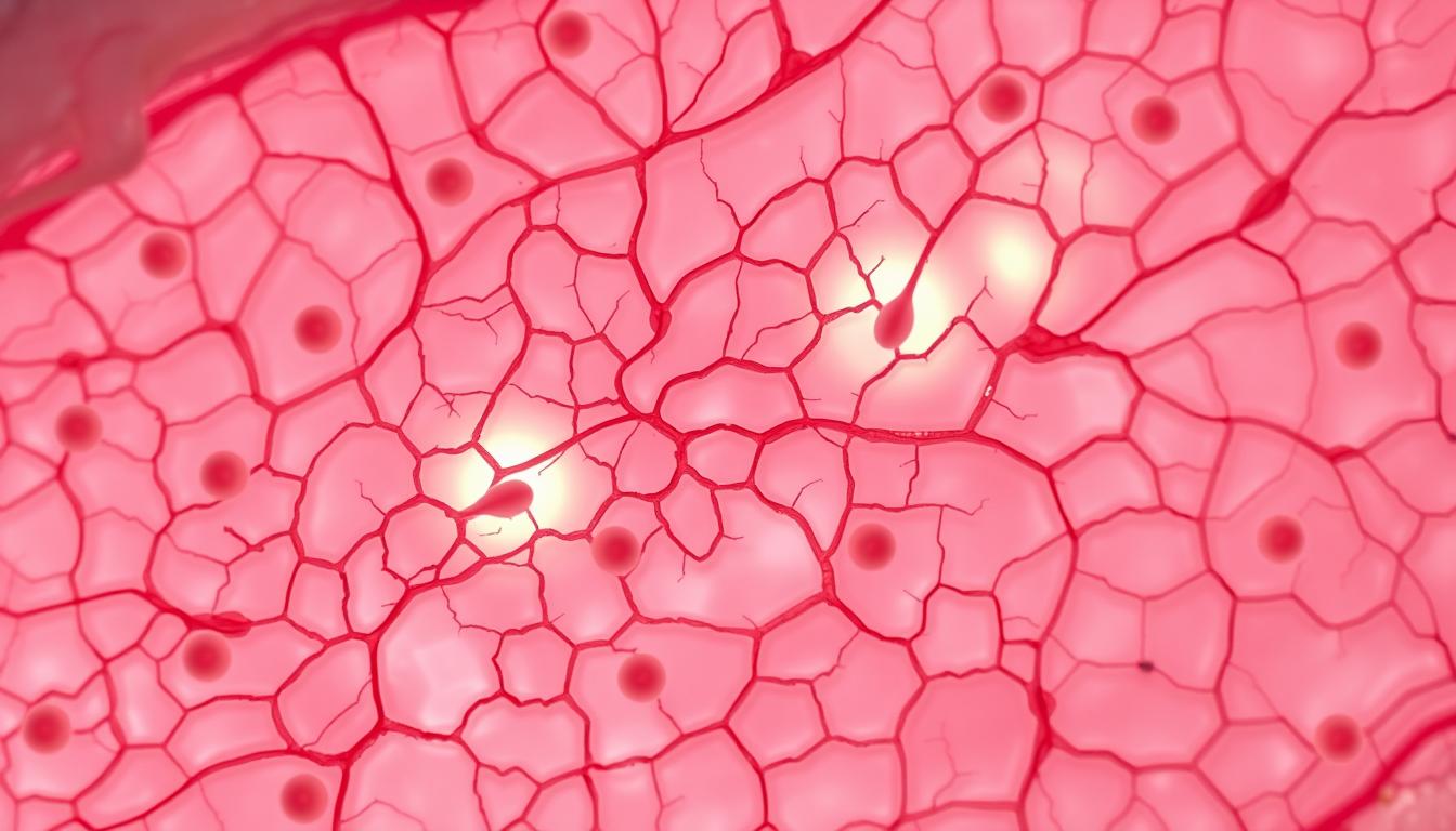
Have you ever wondered what keeps your skin firm, smooth, and resilient? The answer lies deep within the dermis, where specialized cells work tirelessly to maintain its structure. These unsung heroes are essential for both youthful vibrancy and long-term resilience.
Known as the architects of the skin, these cells produce collagen and elastin—proteins that give the skin its strength and elasticity. They also regulate the extracellular matrix, ensuring proper hydration and support. Over time, their activity slows, leading to visible signs of aging.
Understanding their function helps explain why treatments like microneedling and regenerative therapies can rejuvenate the skin. By stimulating these cells, it’s possible to encourage repair and maintain a healthier complexion.
Key Takeaways
- Specialized cells in the dermis maintain skin structure and elasticity.
- Collagen and elastin production is vital for firm, youthful skin.
- The extracellular matrix relies on these cells for hydration and support.
- Aging reduces their activity, contributing to wrinkles and sagging.
- Certain treatments can stimulate them for better skin repair.
Understanding Fibroblasts and Their Structure
Deep within the skin’s layers, specialized cells shape its strength and flexibility. These spindle-shaped builders craft the scaffolding that keeps skin firm and elastic. Their unique design allows them to interact seamlessly with surrounding tissues.
Anatomical Features of Fibroblasts
These cells stand out with long, branching extensions and large, flattened nuclei. Their spindle shape lets them stretch across the extracellular matrix (ECM), weaving collagen and elastin into place. Inside, abundant organelles fuel relentless protein production.
How Fibroblasts Differ from Other Skin Cells
Unlike keratinocytes or melanocytes, fibroblasts excel at ECM synthesis. Key distinctions include:
- Shape: Spindle-like vs. round or cuboidal.
- Function: ECM construction vs. pigment or barrier protein creation.
- Communication: Membrane receptors detect mechanical stress, guiding repair.
This versatility makes them indispensable for wound healing and structural support.
The Critical Role of Fibroblasts in Skin Health
Healthy skin relies on a hidden network of proteins and hydration. Specialized cells craft this foundation, ensuring elasticity and moisture. Their work directly impacts the skin’s youthful appearance.
Fibroblasts and Skin Elasticity
Collagen production forms the backbone of skin’s strength. These cells weave collagen fibrils into dense fibers, providing structure. Simultaneously, elastin fibers cross-link to create stretch and rebound.
Key processes include:
- Tropoelastin assembly into durable elastin networks.
- Lysyl oxidase enzymes stabilizing fiber connections.
- Growth factors signaling repair during stress.
Their Contribution to Skin Hydration
The extracellular matrix acts like a sponge, holding water via glycosaminoglycans (GAGs). Hyaluronic acid, a GAG, binds 1,000 times its weight in water. This keeps skin plump and reduces fine lines.
| Hydration Factor | Function | Impact on Skin |
|---|---|---|
| Proteoglycans | Form moisture-retaining networks | Enhances firmness |
| Hyaluronic Acid | Binds water molecules | Boosts volume |
| Fibronectin | Strengthens ECM scaffolding | Supports repair |
Over time, reduced extracellular matrix efficiency contributes to skin aging. Protecting these systems helps maintain a radiant complexion.
Fibroblasts – Skincare Role in Collagen and Elastin Production
The secret to youthful skin lies in two powerhouse proteins: collagen and elastin. These structures are meticulously crafted by specialized cells, ensuring skin remains firm and flexible. Their production is a precise biological dance, critical for maintaining resilience.
The Science Behind Collagen Synthesis
Collagen forms a triple helix—three polypeptide chains twisted together. Hydroxyproline and glycosylation stabilize this structure, creating durable fibers. Type I collagen dominates the dermis (80–90%), while Type III supports early development and wound repair.
Key steps in synthesis include:
- Gene transcription for alpha chains.
- Post-translational modifications (e.g., hydroxylation).
- Assembly into fibrils via lysyl oxidase cross-linking.
Elastin’s Role in Skin Rebound and Firmness
Elastin enables skin to stretch up to 200% and snap back. Lysyl oxidase bridges tropoelastin molecules, forming a resilient network. Without this process, skin loses its ability to resist sagging.
Mechanical stress triggers fibroblasts to boost protein output. Over time, annual collagen production declines by 1–1.5%, highlighting the need for protective measures like UV avoidance and nutrient-rich diets.
How Fibroblasts Build the Extracellular Matrix
Beneath the surface, a dynamic framework supports the skin’s structure and resilience. This extracellular matrix (ECM) acts like a blueprint, guiding cell behavior and maintaining tissue integrity. Specialized cells assemble its intricate web of proteins and sugars.
Key Components of the ECM
The ECM is a mix of collagens, elastin, and glycosaminoglycans (GAGs). Collagens—especially Type I—form rigid fibers for strength. Elastin provides stretch, while GAGs like hyaluronic acid trap moisture.
Essential ECM elements include:
- Collagens (11+ types): Type I dominates skin; FACIT collagens stabilize networks.
- Elastin: Enables recoil after stretching.
- Proteoglycans: Bind water, cushioning tissues.
| ECM Component | Function | Impact |
|---|---|---|
| Type I Collagen | Provides tensile strength | Prevents sagging |
| Elastin | Allows stretch and rebound | Maintains elasticity |
| Hyaluronic Acid | Retains water | Plumps skin |
Interaction with ECM Proteins
Cells communicate with the ECM via integrins—membrane receptors detecting mechanical stress. This mechano-transduction triggers signaling pathways (e.g., MAPK, Akt) to adjust activity.
Feedback loops are critical. Stiffer ECM boosts collagen production, while MMPs (matrix metalloproteinases) break down old fibers. Aging disrupts this balance, accelerating ECM degradation.
The Connection Between Fibroblasts and Wound Healing
When skin is injured, a precise biological response kicks in to repair the damage. Specialized cells orchestrate this process, ensuring tissues regain strength and flexibility. Their activity peaks within 72 hours, marking the start of a structured recovery phase.
Stages of Fibroblast Activation in Repair
After injury, these cells migrate to the site, initiating proliferation. Key phases include:
- Inflammation: Growth factors like TGF-β signal repair.
- Proliferation: Type III collagen dominates early, forming a temporary scaffold.
- Remodeling: Collagen matures to Type I, strengthening the tissue.
Chronic wounds stall in inflammation due to cellular senescence, delaying healing.
Scar Formation and Fibroblast Activity
Myofibroblasts—activated by mechanical stress—contract the wound, reducing its size. Overactive cells can cause hypertrophic scars, where excess collagen accumulates. Factors like TGF-β imbalance or CD26+ cell activity drive this process.
In diabetic ulcers, impaired growth factors and ECM production hinder recovery. Therapies targeting these pathways aim to restart stalled wound healing.
Fibroblasts and the Aging Process
Aging skin isn’t just about wrinkles—it’s a cellular slowdown with visible consequences. Over time, the activity of key cells drops sharply. By age 80, their population shrinks by 50%, and the extracellular matrix (ECM) renews 75% slower. This decline weakens the skin’s scaffolding, leading to thinning and sagging.
How Aging Affects Cell Function
Replicative senescence—a state where cells stop dividing—plays a major role. After 40–50 divisions, telomeres (protective DNA caps) shorten, halting collagen synthesis. External factors like UV rays accelerate this process, triggering apoptosis (programmed cell death).
Glycation also damages ECM proteins. Sugar molecules bind to collagen, forming stiff cross-links. This reduces elasticity, creating a leathery texture. Key changes include:
- Skin aging markers: Thinned dermis, fragmented elastin.
- Reduced activity of growth factors like TGF-β.
- Fewer receptors for repair signals.
The Link Between Cellular Decline and Wrinkles
Wrinkles form as the elastic network degrades. Without enough support, skin folds deepen. UV exposure worsens this by increasing matrix metalloproteinases (MMPs)—enzymes that break down ECM proteins.
Chronic inflammation from aging or pollution further disrupts balance. Treatments like retinoids or lasers aim to reactivate remaining cells, but prevention—like sunscreen—is critical.
Dermal Fibroblasts vs. Other Skin Cell Types
Skin contains various cell types, each with distinct functions. While fibroblasts build structural support, keratinocytes form the outer barrier. Their differences define how skin stays resilient and protected.
Keratinocytes renew every 28 days, shedding old layers. Fibroblasts, however, live longer and focus on collagen production. This lifecycle contrast ensures both renewal and structural integrity.
ECM output varies too. Fibroblasts produce collagen and elastin, while keratinocytes contribute to the basement membrane. Together, they create a balanced framework for skin strength and hydration.
Immune signaling differs sharply. Fibroblasts interact with immune cells during injury, releasing cytokines. Keratinocytes prioritize barrier defense, activating antimicrobial peptides.
Dermal organization matters. Papillary fibroblasts (upper dermis) support fine structures, while reticular types (deeper layer) anchor thick collagen bundles. This spatial split optimizes skin function.
Stem cell niches rely on fibroblast cues. They secrete laminin and fibronectin, guiding stem cell behavior. Keratinocytes, meanwhile, depend on these signals for proper differentiation.
The Impact of UV Radiation on Fibroblasts
UV rays don’t just tan the skin—they alter its fundamental support system. UVA and UVB penetrate different layers, disrupting collagen production and accelerating aging. UVB alone reduces procollagen I mRNA by 60%, primarily through MMP-1 activation.
How UVA and UVB Damage Skin Differently
UVA reaches the dermis, generating reactive oxygen species (ROS) that cause oxidative DNA damage. UVB targets the epidermis and upper dermis, triggering sunburn cells and cytokine release. Both contribute to photoaging but through distinct pathways.
| UV Type | Penetration Depth | Primary Damage |
|---|---|---|
| UVA | Dermis | ROS, elastosis, MMP upregulation |
| UVB | Epidermis/Upper Dermis | DNA mutations, collagen breakdown |
The Role of ROS and MMPs in Photoaging
ROS like 8-OHdG mark oxidative stress, impairing activity and proliferation. UV exposure also spikes MMP-1, which degrades collagen fibers. Over time, this leads to solar elastosis—abnormal ECM structures that weaken elasticity.
Chronic UV exposure worsens the damage. Studies show UVB increases elastase activity, fragmenting elastic fibers. Combined with reduced collagen synthesis, this creates a visible loss of firmness.
How Lifestyle Choices Influence Fibroblast Health
Daily habits shape skin health more than most realize. From diet to pollution exposure, external factors directly impact the cells responsible for structural support. Optimizing these choices can enhance collagen synthesis and slow aging.
Diet and Nutrient Support
Essential amino acids like glycine and proline fuel collagen production. Vitamin C plays a critical role, enabling hydroxylation—a process that stabilizes collagen fibers. Without it, collagen weakens, leading to fragile skin.
Key nutrients for growth factors include:
- Vitamin C: Supports hydroxylation of proline/lysine.
- Zinc: Co-factor for collagenase enzymes.
- Omega-3s: Reduce inflammation, protecting ECM integrity.
| Nutrient | Function | Food Sources |
|---|---|---|
| Vitamin C | Collagen stabilization | Citrus, bell peppers |
| Glycine | Collagen synthesis | Bone broth, spinach |
| Copper | Elastin cross-linking | Nuts, shellfish |
Note: A balanced diet ensures optimal number of collagen-building blocks.
Smoking and Pollution Effects
Nicotine triggers vasoconstriction, slashing blood flow to skin cells by 40%. This starves them of oxygen, crippling repair activity. Meanwhile, PM2.5 pollutants generate oxidative stress, degrading collagen and elastin.
Alcohol worsens hydration by disrupting hyaluronic acid production. Chronic exposure thins the ECM, accelerating wrinkles. Antioxidants like vitamin E can counteract some damage by neutralizing free radicals.
Protective measures include:
- Antioxidant serums (vitamin C/E).
- Air purifiers to reduce PM2.5 exposure.
- Smoking cessation programs.
Medical Treatments Targeting Fibroblasts
Modern dermatology harnesses advanced techniques to revitalize skin at a cellular level. These methods activate natural repair activity, boosting collagen and elastin production for firmer, younger-looking skin.
Laser Therapy and Cellular Stimulation
Fractional lasers, like the 1540nm wavelength, create controlled micro-injuries. This triggers thermal shock, increasing proliferation and collagen I by 300%. Two primary methods exist:
- Ablative: Removes thin skin layers (e.g., CO2 lasers). Ideal for deep wrinkles.
- Subablative: Heats tissue without damage (e.g., Er:YAG). Less downtime.
Both enhance growth factor release, accelerating repair. Studies show combined treatments yield 40% better results than standalone options.
Platelet-Rich Plasma (PRP) Applications
PRP harnesses the patient’s blood, concentrating platelets rich in healing proteins. Key growth factors include:
| Growth Factor | Function |
|---|---|
| PDGF | Stem cell recruitment, collagen synthesis |
| TGF-β | ECM remodeling, reduces scarring |
| VEGF | Boosts blood flow, nutrient delivery |
Autologous PRP (self-donated) minimizes rejection risks. When paired with lasers, it amplifies activity, reducing recovery time by 50%.
Note: Clinical trials highlight PRP’s role in improving skin texture and elasticity within 6–8 weeks.
Innovative Anti-Aging Therapies Involving Fibroblasts
Cutting-edge science is unlocking new ways to combat aging at the cellular level. Researchers now target the proliferation and activity of key cells, offering lasting rejuvenation. Two groundbreaking methods lead the charge: autologous transplants and stem cell therapies.
Autologous Fibroblast Transplants
This approach uses a patient’s own cells to rebuild collagen networks. Clinicians harvest a small number of cells, culture them in labs, and reinject them into targeted areas. Studies show an 82% reduction in wrinkles after 12 months.
Critical steps include:
- Harvesting: A 3mm skin biopsy from behind the ear.
- Culture: Cells multiply over 6–8 weeks in growth medium.
- Implantation: Optimized density ensures even ECM integration.
| Therapy | Advantage | Outcome Timeline |
|---|---|---|
| Autologous Transplant | No rejection risk | Visible at 3 months |
| PRP + Stem Cells | Enhanced proliferation | Improvement by 6 weeks |
Stem Cell Research and Future Directions
Mesenchymal stem cells (MSCs) differentiate into collagen-producing cells. Their exosomes deliver growth factors, boosting repair. Combined with tissue formation cues, they’re reshaping anti-aging development.
Emerging technologies include:
- 3D Bioprinting: Layers cells into structured ECM mimics.
- Exosome Therapy: Isolates signaling vesicles for targeted renewal.
“MSCs increase collagen synthesis by 200% in hyaluronic acid scaffolds.” — Altman et al, 2010
Fibroblasts in Hair Follicle Health
Hair vitality depends on more than just the strands themselves. Beneath the surface, specialized cells play a crucial part in maintaining healthy hair cycles. These cells support follicle structure and regulate key phases of growth.
Their Role in Hair Growth Cycles
Dermal papilla cells produce insulin-like growth factor-1 (IGF-1), which helps extend the anagen (active) phase. This activity ensures continuous hair production and thickness. Key interactions include:
- ECM remodeling: Collagen and laminin provide structural support.
- TGF-β1 signaling: Initiates catagen (transition) to reset the cycle.
- Nutrient delivery: Blood vessels supply oxygen and proteins for follicle development.
Fibroblasts and Hair Thinning
Reduced cell activity disrupts the balance between growth and shedding. Androgen receptors in dermal papilla cells can trigger miniaturization, leading to thinner strands. Prevention strategies focus on:
| Factor | Impact | Solution |
|---|---|---|
| IGF-1 Decline | Shorter anagen phase | Peptide therapies |
| Oxidative Stress | ECM degradation | Antioxidant serums |
| Androgen Sensitivity | Follicle shrinkage | 5α-reductase inhibitors |
“Fibroblast-derived growth factors maintain 78% of follicular integrity during aging.” — Journal of Dermatological Science
Common Disorders Linked to Fibroblast Dysfunction
Certain skin conditions stem from disruptions in cellular repair mechanisms. When these specialized cells malfunction, they create either excessive or insufficient structural proteins. This imbalance leads to two broad disorders: fibrotic (overproduction) and atrophic (underproduction).
Fibrotic conditions like scleroderma show 300% higher collagen output. The skin thickens and hardens, restricting movement. Atrophic cases involve tissue thinning, often seen in aging or genetic conditions.
Keloids demonstrate dysregulated proliferation. After injury, these cells overproduce collagen, forming raised scars beyond the wound area. Key factors include:
- Abnormal TGF-β signaling
- Reduced apoptosis (cell death)
- Excessive immune cells at the site
Autoimmune attacks target these cells in conditions like lupus. Antibodies mistake them for threats, triggering destructive cytokines. This causes chronic inflammation and ECM degradation.
| Disorder Type | Genetic Component | Primary Symptom |
|---|---|---|
| Ehlers-Danlos | COL5A1 mutation | Hyperelastic skin |
| Werner Syndrome | RECQL2 defect | Premature aging |
Cellular senescence worsens many disorders. Aged cells release SASP (senescence-associated secretory phenotype), including MMPs that break down collagen. ROS accumulation accelerates this process, creating a destructive cycle.
“SASP factors increase inflammation markers by 400% in chronic wounds.” — Journal of Investigative Dermatology
Debunking Myths About Fibroblasts in Skincare
The beauty industry is flooded with bold claims about collagen-boosting products—let’s separate fact from fiction. Many treatments promise dramatic results, but science often tells a different story.
Topical collagen cannot penetrate the dermis. Despite creams claiming to “feed” the skin with collagen, molecules are too large to pass the epidermal barrier. Only internal synthesis by cells creates new structural proteins.
Stem cell serums don’t contain live cells—just their growth factors. These proteins can stimulate activity but won’t “convert” into new cells. Clinical benefits come from consistent use over weeks, not instant transformation.
So-called “activation” creams rarely influence cellular processes directly. Most rely on indirect mechanisms like hydration or mild irritation to trigger natural responses. True activation requires deeper interventions like lasers or microneedling.
“Growth factors in serums are 40,000 Daltons—too large for absorption but effective at signaling surface receptors.” — Dermatology Research Review
Temperature therapies (hot/cold tools) provide temporary tightening only. They don’t boost collagen production long-term. Studies show effects last under 24 hours without structural changes.
- Growth factor stability: Most degrade within 3 months if not refrigerated.
- Retinoids: Thin the epidermis but thicken the dermis over time.
- Peptides: Signal cells but can’t replace professional treatments.
Understanding these limits helps choose effective solutions. Science-backed approaches focus on supporting natural cell functions rather than promising impossible results.
How to Support Fibroblast Activity Naturally
Supporting cellular health doesn’t require complex procedures—simple lifestyle changes make a difference. Research shows natural methods can enhance the skin’s repair activity, promoting collagen and elastin production.
Sleep and Skin Regeneration
Eight hours of sleep increases TGF-β1 by 30%, a key growth factor for tissue repair. Deep sleep phases optimize cellular renewal, while deprivation elevates cortisol, slowing the process.
Dietary Boosters
Antioxidant-rich foods combat oxidative stress:
- Vitamin C: Stabilizes collagen fibers (citrus, bell peppers).
- Omega-3s: Reduce inflammatory cytokines (fatty fish, walnuts).
- Polyphenols: Green tea and turmeric enhance proliferation.
Mechanical Stimulation
Exercise increases blood flow, delivering nutrients to skin cells. For targeted results, microneedling creates micro-injuries that trigger repair—boosting collagen number by 200% in studies.
“Omega-3 supplementation reduces MMP-1 levels by 34%, preserving collagen integrity.” — Dermatologic Surgery Journal
Stress and Hydration
Chronic stress elevates cortisol, impairing growth factors. Mindfulness practices like yoga lower stress markers. Hydration matters too—water supports the extracellular matrix, ensuring efficient nutrient transport.
| Strategy | Mechanism | Impact |
|---|---|---|
| Sleep | TGF-β1 increase | 30% faster repair |
| Antioxidants | ROS neutralization | Reduces collagen breakdown |
| Exercise | Blood flow boost | Enhances nutrient delivery |
Conclusion: The Future of Fibroblast Research in Skincare
Science continues to push boundaries in understanding skin’s natural repair mechanisms. Emerging methods, like exosome therapies and AI-driven ECM modeling, redefine anti-aging development.
Gene editing and mitochondrial transfer techniques promise precision in cellular rejuvenation. Stem cell innovations, combined with epigenetic reprogramming, could personalize treatments.
The future lies in synergistic approaches—lasers paired with PRP or autologous transplants. These combinations optimize the repair process, offering longer-lasting results.
As research advances, expect breakthroughs that blend biology with technology, transforming how we maintain youthful skin.
FAQ
What do fibroblasts do in the skin?
These cells produce collagen, elastin, and other proteins that maintain skin structure, hydration, and elasticity. They also help repair damage and support wound healing.
How do fibroblasts affect aging skin?
As people age, these cells slow down, reducing collagen and elastin production. This leads to wrinkles, thinning skin, and slower healing.
Can you boost fibroblast activity?
Yes. Treatments like laser therapy, PRP, and a nutrient-rich diet can stimulate these cells. Avoiding UV exposure and smoking also helps maintain their function.
What’s the difference between fibroblasts and keratinocytes?
Fibroblasts build the skin’s support structure, while keratinocytes form the outer protective layer. Both are essential but have distinct roles.
Do fibroblasts play a role in hair health?
Yes. They support hair follicles by regulating growth cycles and maintaining the surrounding tissue. Dysfunction can contribute to thinning or loss.
How does the extracellular matrix (ECM) relate to fibroblasts?
These cells create and organize the ECM, a network of proteins like collagen that gives skin strength and flexibility.
Can stress impact fibroblast function?
Chronic stress may reduce their efficiency, slowing repair and accelerating signs of aging. Managing stress supports healthier skin.
Are there medical conditions linked to fibroblast issues?
Yes. Disorders like scleroderma or keloid scars involve abnormal cell behavior, leading to excessive or insufficient collagen production.




Leave a Comment
Your email address will not be published. Required fields are marked *