
Ever wondered about the tiny builders of our body’s tissues? Fibroblasts are the hidden heroes, crucial for our body’s structure.
Fibroblasts are complex cells with a unique shape. They have long, thin extensions that help them work with tissues. Their flat, irregular shape lets them make important parts of our body’s framework.
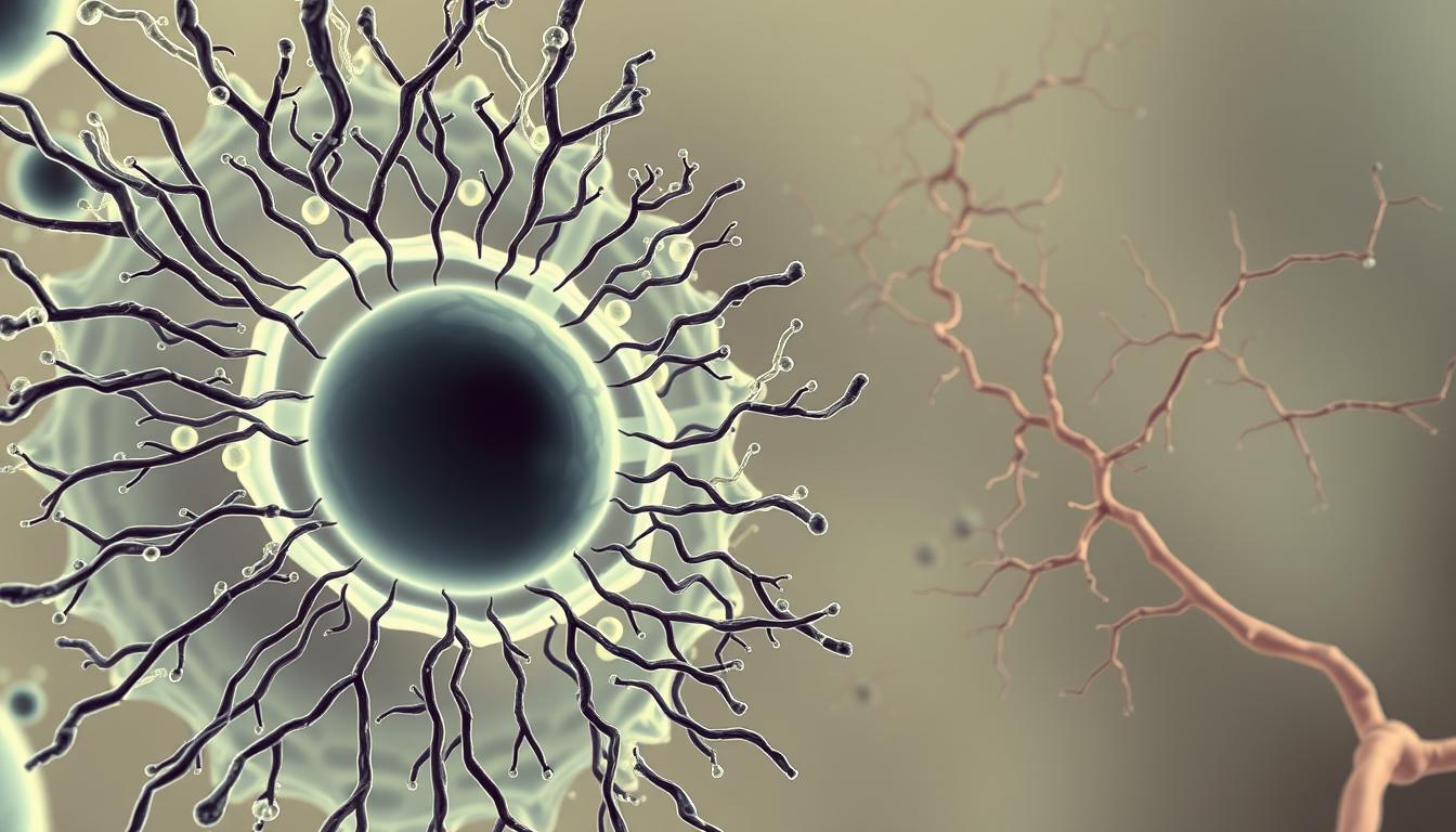
Under a microscope, fibroblasts look like spindles with a big nucleus. They have extensions that help them talk to other cells and move. They make proteins like collagen, which is a big part of our bodies.
Key Takeaways
- Fibroblasts are essential cellular builders in connective tissue
- They possess an elongated, irregular morphological structure
- Capable of producing critical extracellular matrix proteins
- Play a crucial role in tissue repair and maintenance
- Adapt their shape based on tissue requirements
Overview of Fibroblasts in the Human Body
Fibroblasts are amazing cells that help keep our bodies working right. They are key in fixing tissues, handling inflammation, and keeping us healthy. These cells are vital for our body’s health.
Looking into how fibroblasts work shows their incredible skills. They can do many important jobs in different parts of our body.
Definition and Function of Fibroblasts
Fibroblasts are special cells in our connective tissue. They change shape to meet our body’s needs. Their main jobs are:
- Creating the stuff outside our cells
- Helping our tissues stay strong
- Fixing wounds
- Controlling inflammation
“Fibroblasts are the unsung heroes of tissue maintenance, continuously working to preserve our body’s structural integrity.”
Importance in Tissue Repair and Maintenance
Studies show how important fibroblasts are for our tissues. They make a lot of collagen, which is a big part of our skin. This shows how crucial they are for our skin’s health.
Fibroblasts are different in different parts of our body. But, cells from the same area are more alike. This means they have a special role based on where they are.
These cells keep changing the stuff around our cells. They adjust to what our tissues need. Their role in keeping our body balanced is huge.
Structural Characteristics of Fibroblasts
Fibroblasts are fascinating cells that keep tissues healthy and strong. They have unique shapes that help us understand how tissues work.
Size and Shape Considerations
Fibroblasts look flat and long, with extensions that branch out. In a microscope, they are 20 to 50 micrometers long. Their shape is like a spindle.
- Flat, elongated cellular structure
- Branch-like cellular extensions
- Variable size depending on tissue location
Cytoplasmic Features
Fibroblasts have a big nucleus and lots of organelles. This shows they are very active. They make proteins to build the tissue’s framework.
“Fibroblasts are the architects of connective tissue, constantly reshaping and maintaining cellular environments.” – Cellular Biology Research
These cells are very flexible. Their shape lets them meet different needs in different tissues.
Nucleus and Its Role in Fibroblasts
Learning about the nucleus of fibroblasts gives us key insights into how cells work and their genetic processes. The nucleus acts as the main control center, guiding important cell activities with great accuracy.

The structure and genes of fibroblasts are closely tied to their nucleus. When scientists look at fibroblasts under a microscope, they see unique nuclear traits. These traits help define how the cells behave.
Nucleus Structure
The nucleus of a fibroblast looks like a dark, oval shape in the middle of the cell. It has important parts like:
- Nuclear membrane with selective permeability
- Chromatin with genetic info
- Prominent nucleolus for RNA synthesis
- Nuclear pores for molecular transport
Genetic Functionality
Genetic actions in fibroblast nuclei are key for cell repair and keeping tissues healthy. The nucleus manages vital tasks such as:
- DNA replication
- Gene expression regulation
- Protein synthesis control
- Cellular differentiation signals
“The nucleus is not just a storage unit for genetic material, but an active orchestrator of cellular destiny.” – Cell Biology Research Insights
| Nuclear Feature | Primary Function | Significance |
|---|---|---|
| Chromatin | Genetic Information Storage | Regulates Cell Behavior |
| Nucleolus | RNA Synthesis | Protein Production |
| Nuclear Membrane | Molecular Regulation | Selective Permeability |
Every part of the nucleus has a crucial role in keeping fibroblasts working well. This ensures they can repair and maintain tissues effectively.
Fibroblast Appearance Under Microscopy
Looking into the microscopic world of fibroblasts gives us interesting insights. These cells have unique features that scientists can see with advanced tools.
Light Microscopy Visualization
Using light microscopy, researchers see special shapes of fibroblast cells. Active fibroblasts look like spindle-shaped or stellate cells. They have certain traits:
- Oval or centrally placed round nucleus
- Elongated, thin cellular body
- Plump appearance when metabolically active
Electron Microscopy Insights
Electron microscopy shows more details of fibroblast cells. It reveals complex parts inside the cell:
| Cellular Component | Characteristic |
|---|---|
| Nucleus | Euchromatic with prominent nuclear membrane |
| Endoplasmic Reticulum | Extensive rough ER network |
| Golgi Apparatus | Well-developed and prominent |
“The microscopic world of fibroblasts reveals a complex cellular landscape of remarkable structural intricacy.” – Cellular Biology Research Journal
Fibroblasts show a lot of variety in how they look. Inactive fibroblasts are usually longer and have thin, wavy nuclei. This is especially true in areas with lots of collagen fibers. Their shape changes show how important they are for keeping tissues healthy and fixing them when needed.
Variations in Fibroblast Morphology
Fibroblast appearance changes a lot based on where they are and the body’s health. These cells are very flexible and can look different in each body part. This shows how they adapt to their specific roles.
Fibroblasts Across Different Tissue Types
Scientists have found many types of fibroblasts with unique features:
- Cardiac fibroblasts support the heart.
- Dermal fibroblasts keep the skin healthy.
- Muscular fibroblasts help muscles.
- Pericytes keep blood vessels stable.
Morphological Changes in Disease States
Diseases can change how fibroblasts look. Active fibroblasts are usually plump and spindle-shaped. Inactive fibroblasts are longer and have thin, flat nuclei.
“Fibroblasts are dynamic cells that transform their structure in response to physiological challenges” – Cell Biology Research
Diseases like systemic sclerosis can really change fibroblast shape. Studies show that up to 80% of people with systemic sclerosis see changes in their fibrotic tissues. This shows how complex fibroblast changes can be.
Markers like vimentin, PDGFRA, and CD90 help scientists tell fibroblast types apart. This gives them important clues about how these cells change in different situations.
Fibroblast Staining Techniques
Researchers use special staining methods to study fibroblast cells. These methods help see these cells under a microscope. They give important clues about how cells work and look.

Common Staining Approaches
Many staining techniques help scientists see fibroblasts clearly. They use:
- Immunohistochemical staining
- Vimentin marker identification
- Monoclonal antibody techniques
- Fluorescent labeling methods
Advanced Visualization Strategies
Researchers use special markers to show fibroblast features. Vimentin is a key marker, but it’s not always perfect.
Special monoclonal antibodies like anti-pC, 1B10, and 5B5 give detailed views of fibrous tissues.
Interpretation of Staining Results
Understanding staining results needs a lot of skill. Different antibodies show different parts of fibroblasts. This helps scientists learn about how cells work together and their states.
- TE-7 antibody can detect stromal components across 16 tissue types
- Biotin-conjugated secondary antibodies enhance visualization
- Neutral-buffered formalin fixation preserves tissue integrity
These advanced staining methods let scientists study fibroblast cells in great detail. This opens up new areas for medical research and understanding how cells move and work together.
Comparison with Other Cell Types
Cells in the human body are incredibly diverse. Fibroblasts are unique in the connective tissue world. Knowing about their shape and features helps us see how they differ from other cells.
Fibroblasts vs. Myofibroblasts
Fibroblasts can change a lot. They turn into myofibroblasts when tissue gets damaged. This change gives them stronger contractile powers. The main differences are:
- Myofibroblasts have stronger internal fibers
- They can contract more than regular fibroblasts
- Their cells are stiffer
Distinct Characteristics from Other Cell Types
Looking at fibroblasts, adipocytes, and chondrocytes shows interesting differences:
| Cell Type | Size Range | Primary Function | Unique Feature |
|---|---|---|---|
| Fibroblasts | 15-22 µm | Extracellular matrix production | High adaptability |
| Adipocytes | Up to 120 µm | Fat storage | Large lipid droplets |
| Chondrocytes | 20-30 µm | Cartilage maintenance | Collagen type II production |
Fibroblasts are very flexible. They can adjust to different tissue settings while keeping their main roles.
Fibroblasts are the most adaptable cells in connective tissue. They can change and respond to complex signals.
Changes in Fibroblast Appearance with Age
As we age, our fibroblasts change a lot. This affects how well our tissues heal and grow back. Knowing how fibroblasts look at different ages helps us understand aging better.
Cellular Structural Modifications
Fibroblasts look very different as we get older. Scientists have found important changes that affect how they work:
- There are fewer dermal fibroblasts.
- There’s less variety in cells.
- Stem cells don’t renew as well.
- Genomes become more unstable.
Impact on Healing and Repair
Aging fibroblasts can’t do their job as well. Fibroblast morphology changes mean they can’t help tissues heal as much. In people over 80, there’s about a 35% drop in dermal fibroblast numbers compared to the young.
The ability of fibroblasts to repair and maintain tissue integrity declines with age, reflecting fundamental cellular transformation.
Biological Mechanisms of Age-Related Changes
Important changes with age include:
- More DNA damage.
- Shorter telomeres.
- Less sensitivity to growth factors.
- Changes in how collagen is made.
These changes show how fibroblasts gradually lose their ability to heal. This affects our tissue health and how we look.
Role of Fibroblasts in Connective Tissue
Fibroblasts are key cells that help keep connective tissues strong. They have a special structure that lets them make and manage the extracellular matrix (ECM). This matrix is vital for supporting our bodies.

Looking closely at fibroblasts shows how unique they are. They can make over 3.5 million pro-collagen molecules every day. This shows how much they can do.
Types of Connective Tissue Fibroblasts
Fibroblasts have different roles based on where they are in the body. They include:
- Dermal fibroblasts in skin tissue
- Synovial fibroblasts in joint membranes
- Embryonic fibroblasts with regenerative capabilities
- Specialized fibroblasts in different organ systems
Fibroblasts in Extracellular Matrix Production
Fibroblasts mainly make important ECM proteins like collagen and elastin. These proteins help keep tissues strong and support repair.
“Fibroblasts are the architects of our body’s connective tissues, meticulously constructing and maintaining cellular environments.”
| Fibroblast Function | Key Characteristics |
|---|---|
| ECM Protein Synthesis | Produces collagen, elastin, fibronectin |
| Tissue Repair | Supports wound healing processes |
| Cellular Communication | Releases regulatory molecules |
Learning about fibroblasts helps us understand how tissues grow, stay healthy, and can be treated for disorders.
Challenges in Studying Fibroblasts
Studying fibroblasts is complex and requires new methods and technologies. These cells are intricate, making it hard to understand them. Advanced research techniques and visualization methods are needed.
Researchers face many challenges in studying fibroblasts. The main issues include:
- Capturing dynamic cellular interactions
- Identifying subtle morphological changes
- Tracking fibroblast behavior under microscope
- Analyzing heterogeneous cell populations
Limitations of Current Research Techniques
Today’s methods for studying fibroblasts have big limitations. Traditional imaging struggles to show the detailed interactions and states of fibroblasts.
| Research Technique | Current Limitations | Potential Improvements |
|---|---|---|
| Light Microscopy | Low resolution | Advanced optical technologies |
| RNA Sequencing | Limited spatial context | Spatial transcriptomics |
| Immunofluorescence | Static snapshots | Live-cell imaging techniques |
Future Directions in Fibroblast Research
New research strategies are on the horizon. Cutting-edge technologies like single-cell RNA sequencing and advanced microscopy are changing how we understand fibroblasts.
“The future of fibroblast research lies in integrated, multidimensional approaches that combine genetic, molecular, and imaging techniques.” – Contemporary Cell Biology Research
Researchers look forward to big improvements in:
- High-resolution imaging technologies
- Computational modeling of cellular interactions
- Epigenetic profiling techniques
- Functional genomics approaches
These new methods will help us better understand fibroblast behaviors. They could lead to new treatments for many diseases.
Clinical Significance of Fibroblasts
Fibroblasts are vital for our health, keeping tissues in good shape and fixing them when needed. They have special abilities that help them deal with different health challenges.
Learning about fibroblasts shows how important they are in healing wounds and growing new tissue.
Fibroblasts and Wound Healing
Wound healing is a complex process where fibroblasts play a big role. They do several important things:
- They make proteins that help build the tissue’s framework.
- They help rebuild the tissue.
- They help cells move around.
- They help control the body’s response to injury.
“Fibroblasts are the architects of tissue repair, constructing and remodeling damaged structures with remarkable precision.”
Implications in Fibrosis
Fibroblasts can cause problems in diseases like fibrosis. They can make tissues too stiff and scarred.
Studies show fibroblasts are key in many health areas:
- They help manage respiratory diseases.
- They interact with cancer cells.
- They are important in wound healing.
- They help in regenerative medicine.
Because of their many roles, fibroblasts are a focus for new treatments. This could lead to better care in many medical fields.
Conclusion: Understanding Fibroblast Structure
Fibroblasts are key players in human biology. They are found in connective tissue and help keep tissues strong. They also play a big role in healing wounds. Studies show that fibroblasts in different organs have unique structures.
Fibroblasts are not just simple cells. They make proteins that hold tissues together. They can change and adapt to many challenges, like healing wounds or growing new tissue. This shows how important they are for keeping cells healthy.
Summary of Key Features
Fibroblasts are very versatile. They come in different types, like those found in the heart, skin, and muscles. Each type is suited for its specific environment. This makes them very interesting for medical research, especially in understanding how tissues heal and change with age.
Future Implications for Research and Medicine
Studying fibroblasts could lead to big breakthroughs in health. They might help in treating heart disease, inflammation, and cancer. By learning more about how fibroblasts work, scientists could find new ways to fix damaged tissues and create personalized treatments.
FAQ
What exactly are fibroblasts?
Fibroblasts are key cells in connective tissue. They help keep tissues strong and intact. They make proteins like collagen that support our bodies.
How do fibroblasts appear under a microscope?
Under a microscope, fibroblasts look like long, spindle-shaped cells. They have a big nucleus and long arms. These arms help them connect with other cells and the matrix.
What is the primary function of fibroblasts in the human body?
Fibroblasts are vital for fixing and keeping tissues healthy. They make proteins like collagen and fibronectin. They help heal wounds and support tissue growth.
Do fibroblasts change with age?
Yes, fibroblasts change as we age. They work less, grow slower, and make less matrix. These changes affect how well they repair tissues.
How do fibroblasts differ from other cell types?
Fibroblasts are different from cells like myofibroblasts or adipocytes. They are long and flexible. This shape lets them move and work in the matrix.
Can fibroblasts be found in different types of tissues?
Yes, fibroblasts are in almost all connective tissues. They are in skin, tendons, ligaments, and organs. Their look and function vary by tissue.
What techniques are used to study fibroblast structure?
Scientists use light and electron microscopy, and special stains like immunofluorescence. These help them see and study fibroblasts’ structure and how they work.
Are fibroblasts important in medical research?
Fibroblasts are very important in medical research. They help us understand how to heal wounds, grow tissues, and treat fibrosis. This is key for many connective tissue disorders.


Leave a Comment
Your email address will not be published. Required fields are marked *