
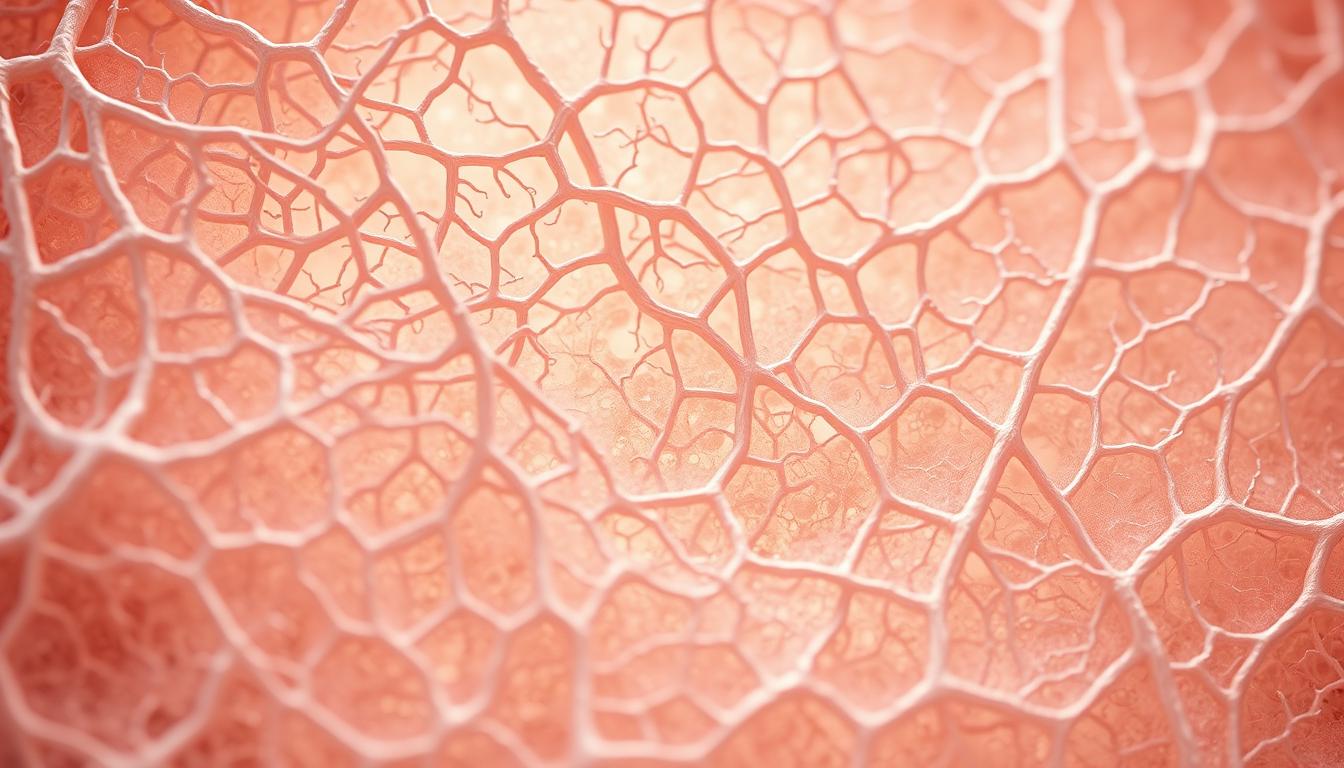
Ever wondered how your skin stays firm or your tendons remain strong? The secret lies in two critical components: collagen fibers and fibroblasts. While one acts as the body’s structural glue, the other tirelessly works to produce and maintain it.
Collagen fibers are sturdy proteins that form the extracellular matrix, giving tissues their strength. In contrast, fibroblasts are active cells that synthesize these fibers, ensuring tissues stay resilient. Together, they play a vital role in healing, growth, and overall health.
Key Takeaways
- Collagen fibers provide structural support to tissues.
- Fibroblasts are the cells responsible for producing collagen.
- Collagen’s triple-helix structure ensures durability.
- Fibroblasts actively repair and remodel tissues.
- Type I collagen is the most abundant in the human body.
Understanding Fibroblasts and Collagen: Core Definitions
Behind every resilient tissue lies a dynamic interplay of cells and structural proteins. These elements ensure our body’s framework remains sturdy yet adaptable. Let’s break down their core roles.
What Are Fibroblasts?
First identified in 1858, fibroblasts are spindle-shaped cells found in connective tissues. They produce critical components like collagen, elastin, and fibronectin. Their extensive endoplasmic reticulum enables efficient protein synthesis.
These cells adapt to needs—transforming into myofibroblasts during wound healing or adipocytes in fat storage. They maintain tissue homeostasis by balancing matrix production and breakdown.
What Are Collagen Fibers?
Collagen is the body’s most abundant protein, forming rope-like fibers for tensile strength. Synthesized by fibroblasts, its triple-helix structure resists stretching. Vitamin C is essential for its production.
In skin, collagen types I and III dominate, providing elasticity and support. These fibers also guide cell migration during repair processes.
| Feature | Fibroblasts | Collagen Fibers |
|---|---|---|
| Nature | Living cells | Non-living protein structures |
| Primary Role | ECM synthesis and repair | Structural support |
| Key Component | Endoplasmic reticulum | Triple-helix amino acid chains |
Fibroblasts vs. Collagen: Key Structural and Functional Differences
Structural stability in the body comes from a balance of living cells and protein networks. Fibroblasts and collagen work together yet differ fundamentally in their roles and behaviors.
Fibroblasts are dynamic cells marked by PDGFRA/VIM proteins. They actively synthesize proteins, requiring energy for ribosomal processes. In contrast, collagen fibrils (1–2μm in gels) are passive structures formed enzymatically.
Their lifespans differ sharply. Fibroblasts renew through division, while collagen fibers persist longer but rely on cellular activity for remodeling.
Core Contrasts
| Feature | Fibroblasts | Collagen |
|---|---|---|
| Nature | Living, metabolically active | Non-living, structural |
| Primary Function | ECM production, repair | Tensile strength |
| Synthesis | Ribosomal protein assembly | Enzymatic cross-linking |
| Disease Link | Fibrosis (overactivation) | Genetic disorders (e.g., EDS) |
Mechanically, fibroblasts contract tissues during healing, while collagen resists stretching. Their functions complement each other in maintaining tissues.
For deeper insights into fibroblast activity, explore their key products and functions.
The Role of Fibroblasts in Collagen Production
Tissue resilience stems from a carefully orchestrated protein synthesis process. Specialized cells build and regulate the structural proteins that keep tissues intact. Central to this is collagen production, a multi-step feat of molecular engineering.
How Fibroblasts Synthesize Collagen
The process begins in ribosomes, where alpha chains are translated. These chains then enter the endoplasmic reticulum (ER) for critical modifications:
- Prolyl hydroxylation: Enzymes add hydroxyl groups to proline residues, stabilizing the future triple helix.
- Glycosylation: Sugar molecules attach to hydroxylysine, aiding fiber stability.
In the Golgi apparatus, three alpha chains twist into a triple helix—collagen’s signature structure. Vesicles transport this procollagen to the cell membrane for secretion.
Regulation of Collagen Secretion
Growth factors like TGF-β1 dramatically boost output. Studies show it increases type I collagen production by 5.8-fold. Other regulators include:
| Factor | Effect | Mechanism |
|---|---|---|
| TGF-β1 | Stimulates synthesis | Activates fibroblast genes |
| PDGF | Enhances proliferation | Signals via tyrosine kinases |
| Vitamin C | Cofactor for hydroxylation | Supports ER enzymes |
External stiffness also influences output. Fibroblasts in rigid environments (e.g., scar tissue) produce more collagen. Learn how these fibroblast functions adapt to tissue demands.
Types of Collagen Produced by Fibroblasts
The human body relies on distinct protein structures to maintain tissue integrity. Among these, type collagen variants like I and III play specialized roles in structural support and flexibility.
Type I Collagen: The Most Abundant Form
Type I dominates connective tissues, making up 90% of skin collagen. Its fibrillar structure provides unmatched tensile strength. Key features include:
- Found in skin, bones, and tendons.
- Mature cross-links resist enzymatic degradation.
- Mutations in COL1A1/COL1A2 genes cause osteogenesis imperfecta (“brittle bone disease”).
“Type I collagen’s rigidity is why it’s the scaffold for load-bearing tissues.”
Type III Collagen and Its Specialized Roles
Often paired with Type I, type III forms reticular fibers in elastic tissues like blood vessels and lungs. It increases 14.9-fold in pulmonary fibrosis, indicating its role in pathological remodeling.
| Feature | Type I | Type III |
|---|---|---|
| Primary Location | Skin, bones | Blood vessels, lungs |
| Clinical Link | Osteogenesis imperfecta | Fibrotic disorders |
| Structural Role | Rigid support | Elasticity |
While Type I ensures durability, Type III offers flexibility—both critical for healthy tissues.
Extracellular Matrix: Where Fibroblasts and Collagen Interact
Connective tissues owe their resilience to intricate molecular teamwork. The extracellular matrix (ECM) serves as the stage for this collaboration, housing over 300 core proteins that guide cellular behavior. Here, structural molecules and cells communicate to maintain tissue function.
Fibroblasts constantly sense and remodel the ECM. Through integrin receptors, they detect mechanical cues like stiffness, triggering pathways that alter protein production. This feedback loop ensures tissues adapt to stress or injury.
Key ECM components include:
- Proteoglycans: Hydrate the matrix and buffer forces.
- Fibronectin: Anchors cells to collagen networks.
- Matrix metalloproteinases (MMPs): Enzymes that break down excess proteins.
In rigid environments, fibroblasts differentiate into myofibroblasts, amplifying ECM deposition. This response aids wound healing but can spiral into fibrosis if unchecked. Studies link TGF-β1 signaling to such pathological remodeling.
“The ECM isn’t just a scaffold—it’s a dynamic instructor that shapes cell fate.”
Cancer exploits these mechanisms. Tumors stiffen the connective tissue, hijacking fibroblast activity to spread. Researchers now target ECM interactions to block metastasis.
Fibroblast Heterogeneity Across Tissues
Not all structural cells are created equal. Depending on their location, dermal fibroblasts and other specialized variants adapt to meet unique tissue demands. This diversity stems from distinct gene expression patterns and microenvironment cues.
Dermal vs. Pulmonary Fibroblasts
Skin and lung cells exhibit striking functional contrasts. Studies show pulmonary variants migrate 10% faster than their dermal counterparts. Their gene expression profiles also differ significantly:
- TGF-β1 response: Lung cells show amplified sensitivity to this growth factor.
- ECM output: Skin cells prioritize Type I collagen, while lung cells focus on elastin.
- Positional memory: Dermal variants retain location-specific HOX gene signatures.
These adaptations enable tissue-specific functions. For example, pulmonary cells must accommodate constant airflow changes.
Organ-Specific ECM Adaptations
Each tissue shapes its structural cells differently. Biomarkers like COL6A3 help identify these specialized populations. Consider how heart and bladder fibroblasts differ:
| Tissue | Key ECM Protein | Unique Marker |
|---|---|---|
| Skin | Type I collagen | COL12A1 |
| Lung | Elastin | COL6A5 |
| Heart | Fibronectin | Thbs1 |
Such variations explain why skin heals differently than lung tissue. The tissues essentially “train” their resident cells through mechanical and chemical signals.
“A lung fibroblast transplanted to skin would still try to build lung matrix—that’s how deep this specialization runs.”
Fibroblasts – Collagen Difference in Wound Healing
When injuries occur, the body initiates a precise repair process involving specialized cells and structural proteins. This coordinated effort ensures tissues regain strength and function. Central to this role are two key players with distinct contributions.
Fibroblast Activation and Myofibroblast Differentiation
Initially dormant cells transform into active builders during wound healing. Growth factors like PDGF trigger this shift, turning them into synthetic powerhouses. They begin producing ECM components at accelerated rates.
A critical transition occurs when these cells develop α-SMA stress fibers. This change marks their evolution into myofibroblasts—specialized contractile cells. Their role includes:
- Pulling wound edges together through contraction
- Depositing new matrix proteins
- Regulating inflammation levels
“Myofibroblasts are nature’s stitches—they physically draw damaged tissues back together while rebuilding the structural framework.”
Collagen Remodeling During Tissue Repair
The extracellular matrix undergoes dramatic changes throughout recovery. Early stages feature Type III protein networks—flexible but weak. Over time, these get replaced by stronger Type I formations.
| Healing Phase | Protein Type | Function |
|---|---|---|
| Inflammatory | Fibronectin | Scaffold formation |
| Proliferative | Type III | Initial matrix |
| Remodeling | Type I | Mature strength |
Matrix metalloproteinases carefully remove excess proteins during later stages. This balanced process prevents abnormal scarring while restoring tissue integrity.
Clinical applications leverage this knowledge. Therapies targeting TGF-β1 signaling show promise in reducing pathological scarring. Other approaches aim to optimize the wound healing sequence for better outcomes.
Single-Cell Transcriptomics: Revealing Fibroblast Diversity
Advanced tools expose intricate cellular variations previously invisible to researchers. Single-cell RNA sequencing (scRNA-seq) now maps these differences, uncovering specialized subtypes like *Pi16+/Col15a1+* cells. Such studies highlight how tissues tailor their structural networks.
Technical comparisons between methods like Drop-seq and SmartSeq2 reveal trade-offs. While Drop-seq captures more cells, SmartSeq2 offers deeper gene expression profiles. This precision helps identify rare populations, such as *Pdgfra+/Dpt+* clusters in connective tissues.
Cluster analysis further divides cells into functional groups. Some subsets drive inflammation, while others focus on matrix production. Spatial transcriptomics adds context, showing how these groups organize within tissues.
“ScRNA-seq transforms fibroblasts from generic ‘worker cells’ into nuanced characters with scripted roles in health and disease.”
Pseudotime analysis tracks how cells evolve during activation. Cross-species research confirms some subtypes are conserved, like those in fibrotic lungs. Disease-specific states, such as cancer-associated profiles, are now identifiable.
These breakthroughs redefine research approaches. By decoding cellular diversity, scientists can target precise populations for therapies. The next frontier? Linking these gene expression patterns to functional outcomes in real time.
Collagen Fiber Formation and Assembly
From tiny molecules to robust fibers, collagen transforms through ordered steps. This protein gains strength via hierarchical assembly, ensuring tissues withstand mechanical stress. The process involves two critical phases: fibrillogenesis and cross-linking.
Fibrillogenesis: From Molecules to Fibers
Collagen monomers first aggregate via nucleation-controlled polymerization. Here, small clusters act as templates for larger fibrils. Studies reveal these grow through lateral fusion of microfibrils, creating the classic 67nm banding pattern.
Specialized protein groups regulate this growth. FACIT collagens (e.g., Types IX, XII) limit excessive expansion. Meanwhile, proteoglycans like decorin stabilize fibril surfaces by binding core proteins.
Cross-Linking and Mechanical Strength
Lysyl oxidase (LOX) enzymes finalize fibril strength. They create covalent bonds between hydroxylysine residues, forming durable networks. This collagen production step is vital for tissue integrity—mutations in LOX genes lead to fragile skin and vessels.
| Process | Key Mechanism | Outcome |
|---|---|---|
| Fibrillogenesis | Lateral fusion of microfibrils | 67nm banded fibrils |
| Cross-linking | LOX-mediated covalent bonds | High tensile strength |
“Collagen’s resilience isn’t innate—it’s engineered through precise molecular partnerships.”
These steps ensure fibers adapt to tissue needs. In tendons, tightly packed fibrils resist stretching. In skin, looser networks allow flexibility—all governed by the same collagen production principles.
Pathological Changes: Fibrosis and Scarring
When tissue repair goes wrong, the body’s natural healing process can spiral into harmful overdrive. Fibrotic diseases like systemic sclerosis and pulmonary fibrosis stem from this imbalance, replacing functional tissue with stiff, non-elastic scars. The result? Impaired organ function and chronic pain.
Excessive Collagen Deposition in Disease
In healthy healing, collagen production slows once wounds close. But in fibrosis, signals go awry. Hypoxia triggers HIF-1α, boosting protein synthesis. Autoantibodies further activate cells, creating a vicious cycle. Studies show excessive collagen levels in SSc-ILD rise 8.8-fold (PRO-C1 biomarkers).
Key drivers include:
- ECM stiffness: Abnormal mechanosensing locks cells into overproduction.
- Failed resolution: MMPs can’t break down surplus proteins.
“Fibrosis isn’t just scar tissue—it’s a pathological rewrite of the extracellular matrix.”
Fibroblast Dysregulation in Systemic Sclerosis
Systemic sclerosis (SSc) epitomizes fibroblast dysfunction. Cells ignore stop signals, flooding tissues with ECM proteins. The FDA-approved drug nintedanib now targets this by inhibiting tyrosine kinases.
| Feature | Healthy Healing | Fibrosis |
|---|---|---|
| Collagen Turnover | Balanced | Excessive deposition |
| Cell Activity | Self-limiting | Persistent activation |
| Therapy Example | None needed | Nintedanib |
Research highlights TGF-β1’s role in this dysregulation. It flips fibroblasts into overdrive, stiffening tissues irreversibly. Emerging treatments aim to recalibrate these signals.
3D Models: Studying Fibroblast-Collagen Interactions
Modern research tools allow scientists to explore cellular dynamics in lifelike environments. Advanced 3D models reveal how structural proteins and cells interact under realistic conditions. These studies provide critical insights into tissue repair and disease progression.
Collagen Gel Contraction Assays
Researchers use collagen gels to measure how cells reshape their surroundings. These assays quantify contractile forces, revealing key aspects of cell behavior. Common methods include:
- TGF-β1 dose tests: Tracking response curves to growth factors.
- Inhibitor screening: Testing drugs that block contraction.
- Stiffness calibration: Adjusting matrix density to mimic tissues.
Floating gels (1-2mg/ml) simulate tumor environments, while attached models mirror healthy tissues. This helps pinpoint how mechanical stress alters cellular responses.
Mechanical Stress and Cell Behavior
Cells react differently based on matrix tension. High-stress conditions trigger distinct signaling pathways like Rho kinase. Key findings include:
| Condition | Cell Response | Key Pathway |
|---|---|---|
| Low tension | Dendritic extensions form | Microtubule-dependent |
| High tension | Force transmission increases | PAK1/cofilin1 |
“3D models bridge the gap between petri dishes and living tissues—showing us how cells truly behave under pressure.”
These studies explain why some wounds heal poorly. They also guide therapies targeting cell behavior in fibrotic diseases.
Growth Factors Influencing Fibroblast-Collagen Dynamics
Molecular signals dictate how structural proteins and cells coordinate tissue repair. Growth factors like TGF-β1 and PDGF serve as master regulators, activating precise signaling pathways to balance production and remodeling.
TGF-β1: The Master Regulator
TGF-β1 boosts COL1A1 gene expression 2.2-fold in lung tissues. Its cascade begins with SMAD2/3 phosphorylation, triggering:
- Collagen synthesis: Enhanced ribosomal translation of matrix proteins.
- Fibronectin production: Increased adhesion molecule output (P
- Negative feedback: SMAD7 halts excessive activation.
Drugs like iALK5 inhibit this pathway, reducing protein formation by 50%.
PDGF: Driving Migration and Proliferation
PDGF-AB elevates cell migration by 66%. It activates receptor tyrosine kinases, stimulating:
| Effect | Mechanism |
|---|---|
| Type VI collagen surge | P |
| MAP kinase cross-talk | Synergizes with TGF-β1 signals |
“Targeting PDGF and TGF-β1 pathways simultaneously may reverse fibrotic damage more effectively than single-pathway inhibitors.”
These factors exemplify how cells decode biochemical cues to maintain tissue integrity. Disruptions in their balance underlie diseases like fibrosis, driving therapeutic innovation.
Comparative Analysis: Fibroblasts vs. Other Connective Tissue Cells
Connective tissues host diverse cell populations with specialized roles. While fibroblasts dominate matrix production, other cell types contribute unique functions essential for tissue performance. Understanding these distinctions helps explain how bones, cartilage, and tendons maintain their specific properties.
Recent single-cell studies reveal key contrasts. Pericytes, often confused with fibroblasts, show distinct PDGFRB expression versus fibroblast PDGFRA markers. Myofibroblasts further differentiate by expressing α-SMA, blending muscle-like contraction with synthetic functions.
| Cell Type | Key Feature | Primary Output |
|---|---|---|
| Chondrocyte | Type II collagen specialization | Cartilage matrix |
| Osteoblast | Mineralizing function | Bone tissue |
| Adipocyte | Lipid storage vesicles | Energy reserves |
| Tenocyte | Force transmission adaptations | Tendon strength |
| Pericyte | Vascular support | Capillary stability |
Chondrocytes exemplify tissue-specific adaptation. They exclusively produce Type II collagen—unlike fibroblasts—creating cartilage’s shock-absorbing properties. This specialization allows joints to withstand compression forces during movement.
“Single-cell technologies prove what histology hinted: every connective tissue cell speaks its own molecular language.”
Osteoblasts demonstrate another extreme. These cells deposit hydroxyapatite crystals, transforming soft connective tissue cells into rigid bone. Their activity depends on precise Wnt signaling pathways absent in typical fibroblasts.
Adipocytes and tenocytes show how environment shapes function. Fat cells store energy in lipid droplets, while tendon cells align collagen fibers for mechanical stress. Such adaptations make each cell type irreplaceable in its niche.
Technological Advances in Fibroblast Research
Cutting-edge technologies are reshaping how scientists study cellular behavior. From decoding genetic activity to mapping spatial relationships, these tools reveal unprecedented details about tissue dynamics. Two innovations lead this revolution: single-cell RNA sequencing and advanced bioinformatics.
Single-Cell RNA Sequencing Breakthroughs
Modern platforms like Drop-seq profile thousands of cells simultaneously. This high-throughput approach identifies rare subtypes, such as *Pi16+/Col15a1+* cells in connective tissues. Key enhancements include:
- Cell hashing: Labels cells for multiplexing, boosting experimental efficiency.
- Multiome ATAC-seq: Combines gene expression and chromatin data for holistic analysis.
Unlike traditional methods, snRNA-seq preserves ECM-embedded cells, avoiding dissociation artifacts. This is critical for accurate studies of structural networks.
Bioinformatics and Classification Challenges
Analyzing scRNA-seq data requires robust algorithms. While UMAP and t-SNE visualize clusters, they often distort cell-type relationships. New tools address this by:
| Tool | Advantage |
|---|---|
| SPRING | Maintains global data structure |
| PHATE | Preserves trajectory relationships |
“Spatial transcriptomics bridges molecular data with tissue architecture—showing not just what cells express, but where.”
Integrating these with *bioinformatics* pipelines ensures precise classification. For example, mapping *Pdgfra+/Dpt+* clusters reveals their role in fibrosis progression.
Clinical Implications of Fibroblast-Collagen Research
Recent FDA approvals highlight progress in combating tissue scarring. The 2019 clearance of nintedanib for systemic sclerosis-associated interstitial lung disease (SSc-ILD) marked a turning point. This tyrosine kinase inhibitor reduces disease progression by 43%, showcasing how drug development leverages cellular research.
- Biomarker discovery: PRO-C1 assays now quantify collagen turnover in real time.
- Targeted delivery: Nanoparticles carrying siRNA selectively silence fibrotic genes.
- Senolytic strategies: Dasatinib/quercetin combinations clear aged cells in trials.
The Scar-in-a-Jar model accelerates preclinical testing. This 3D system mimics human fibrosis, allowing rapid screening of 320+ compounds weekly. Researchers prioritize candidates that normalize ECM stiffness without harming healthy tissues.
| Approach | Mechanism | Status |
|---|---|---|
| LOXL2 inhibitors | Block collagen cross-linking | Phase III trials |
| CRISPR-Cas9 editing | Disrupt TGF-β1 receptors | Preclinical |
| ECM-modifying enzymes | Degrade excess fibronectin | FDA Fast Track |
“Tissue-specific drug targeting doubles response rates compared to systemic therapies in SSc-ILD cohorts.”
Emerging drug development pipelines now address organ-specific variations. Pulmonary formulations prioritize elastase inhibition, while dermal therapies focus on MMP-1 activation. These advances reflect deeper understanding of clinical implications across tissue types.
Conclusion
The intricate dance between structural proteins and their cellular architects shapes tissue health. While one provides the body’s framework, the other dynamically builds and remodels it—a partnership critical for healing and disease prevention.
Microenvironmental cues dictate these interactions, from mechanical stiffness to biochemical signals. Emerging 3D models now capture this complexity, offering new ways to study therapeutic targets for fibrosis and scarring.
Unanswered questions remain, particularly about cell heterogeneity across tissues. Interdisciplinary approaches—combining molecular biology, engineering, and clinical research—will drive the next wave of breakthroughs in regenerative medicine.
FAQ
What are fibroblasts and collagen fibers?
Fibroblasts are cells that produce and maintain the extracellular matrix, while collagen fibers are structural proteins providing strength and elasticity to tissues. Both play essential roles in tissue integrity and repair.
How do fibroblasts contribute to collagen production?
These cells synthesize and secrete collagen precursors, which then assemble into fibers. Growth factors like TGF-β1 regulate this process, ensuring proper tissue structure and function.
What types of collagen do fibroblasts produce?
They primarily generate Type I (found in skin and bones) and Type III (common in blood vessels and organs). Each type has distinct mechanical and biological properties.
How do fibroblasts and collagen interact in wound healing?
During repair, activated cells differentiate into myofibroblasts, contracting the wound and depositing new fibers. Proper remodeling ensures functional tissue restoration.
What happens when collagen production becomes excessive?
Overproduction leads to fibrosis, causing stiff, scarred tissues. Conditions like systemic sclerosis result from dysregulated fibroblast activity and abnormal ECM accumulation.
How do researchers study fibroblast-collagen interactions?
Advanced techniques like 3D gel assays and single-cell RNA sequencing reveal how mechanical forces and gene expression influence ECM dynamics in health and disease.
Are fibroblasts the same in all tissues?
No, they exhibit organ-specific variations. For example, dermal cells differ from pulmonary fibroblasts in gene expression and ECM composition, adapting to unique tissue demands.
Why is collagen cross-linking important?
Cross-linking enhances fiber stability and tensile strength. Enzymes like LOX facilitate this process, ensuring tissues withstand mechanical stress without degradation.


Leave a Comment
Your email address will not be published. Required fields are marked *