
Did you know that nearly 50% of deaths in developed nations stem from disorders linked to a single cell type? These versatile cells, essential for tissue repair, can also drive severe medical conditions when malfunctioning.
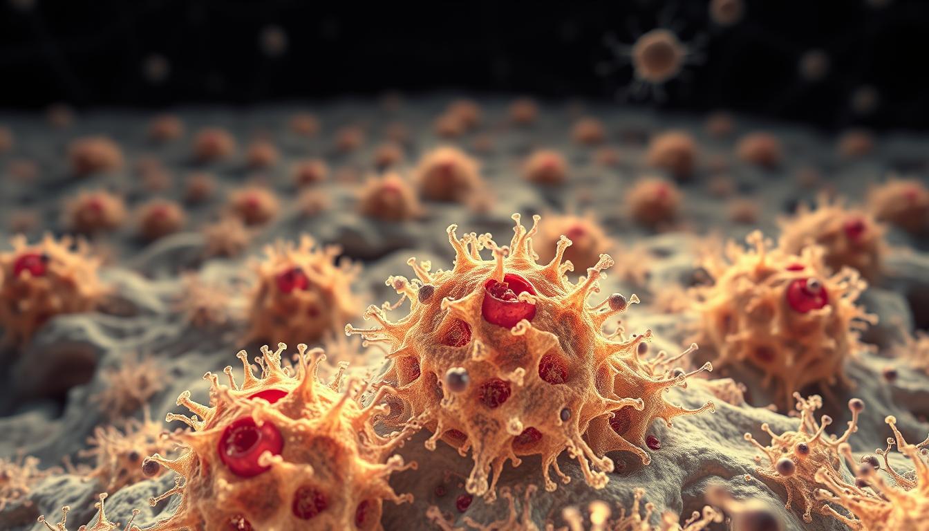
Fibroblasts are mesenchymal cells responsible for maintaining tissue structure and healing wounds. While they play a crucial role in normal bodily functions, their abnormal activity contributes to chronic illnesses. Recent studies highlight their involvement in fibrosis, autoimmune disorders, and even cancer progression.
Advanced techniques like single-cell RNA sequencing reveal surprising diversity among these cells. This discovery helps explain why they influence multiple conditions differently across organs. Understanding their mechanisms could unlock new treatments for previously untreatable diseases.
Key Takeaways
- Fibrosis accounts for half of mortality cases in developed countries
- These cells exhibit organ-specific variations affecting disease development
- Autoimmune conditions and cancer progression involve fibroblast dysfunction
- Cutting-edge research uncovers previously unknown cell heterogeneity
- Historical observations from 1858 still inform modern studies
The Essential Role of Fibroblasts in Tissue Homeostasis
These unsung heroes of the body quietly maintain structural integrity while orchestrating complex cellular communication. Their dual role as builders and messengers makes them indispensable for healthy tissue function.
Extracellular Matrix Synthesis and Remodeling
Specialized cells produce 80% of structural proteins that form the body’s scaffolding. This intricate network includes:
- Collagen fibers for tensile strength
- Elastin proteins for tissue flexibility
- Proteoglycans for hydration and cushioning
Matrix metalloproteinases act as precision tools, constantly reshaping this framework. “The extracellular matrix isn’t static – it’s a dynamic, living structure,” notes a 2021 Cell study. Mechanical forces generated through this remodeling process guide cell behavior and tissue organization.
Fibroblasts as Signaling Niche Cells
Beyond structural support, these cells create microenvironments that regulate neighboring cells. They secrete vital compounds including:
- Growth factors like TGF-β and FGF
- Metabolic regulators
- Positional markers through HOX genes
Wnt and Notch pathways maintain stem cell niches, influencing tissue repair. Research from embryonic chick heart studies reveals how early these signaling patterns establish themselves. Regional specialization explains why skin and heart tissues develop distinct characteristics despite similar building blocks.
The balance between normal function and overactivation proves crucial. While essential for development and healing, excessive activity leads to pathological changes. Understanding these mechanisms opens new therapeutic possibilities for chronic conditions.
Fibroblasts – Related Diseases: An Overview
Over 120 medical conditions trace back to malfunctioning cells that build our body’s framework. Nature Reviews Immunology (2021) confirms their dysregulation spans fibrosis, autoimmune disorders, and cancer. These conditions arise through three core mechanisms:
- Excessive ECM production: Stiffens organs like lungs and liver
- Chronic inflammation: Fuels joint damage in rheumatoid arthritis
- Tumor support: Shields cancers from immune attacks
Acute activation aids wound healing, but chronic signaling spirals into pathology. Single-cell studies reveal specialized subtypes like myofibroblasts and cancer-associated fibroblasts (CAFs) drive disease progression uniquely in each context.
| Condition | Prevalence (per 100,000) | Primary Mechanism |
|---|---|---|
| Rheumatoid Arthritis | 400-1,300 | Synovial fibroblast hyperactivity |
| Systemic Sclerosis | 40-340 | Collagen overproduction |
| Lupus Erythematosus | 20-150 | Immune-fibroblast miscommunication |
Buechler et al. (2021) mapped fibroblast diversity across tissues, uncovering shared inflammatory signatures in RA and bowel disease. Yet early detection remains tough—these cells mask symptoms until irreversible damage occurs.
Their dialogue with immune cells worsens autoimmunity. For example, synovial fibroblasts in arthritis secrete cytokines that activate T cells, creating a vicious cycle. Targeted therapies now aim to interrupt these conversations.
Fibrosis: When Fibroblasts Turn Pathogenic
When healing turns harmful, fibrosis emerges as a silent threat. This condition occurs when excessive scar tissue stiffens organs, disrupting their function. Overactivation of repair mechanisms drives the process, often triggered by chronic injury or inflammation.
How Cells Shift from Repair to Damage
The TGF-β/Smad pathway acts as the master switch. It triggers cells to produce three times more collagen than normal, as shown in a 2021 Cell study. Mechanical stress, like high blood pressure in the heart, further amplifies this response.
Other key players include:
- PDGF receptors: Overexpressed in scleroderma, fueling uncontrolled growth
- Epithelial-mesenchymal transition (EMT): Converts lung cells into collagen-producing machines
- Pericyte conversion: Mouse models reveal blood vessel cells transforming into myofibroblasts
Fibrosis Patterns Across Organs
Lungs and liver show stark contrasts:
| Organ | Key Feature | Driver |
|---|---|---|
| Lung (IPF) | Stiff air sacs | Lipofibroblast transition |
| Liver | Scarred lobes | Activated stellate cells |
| Skin | Thickened patches | PI16+ subset persistence |
Cardiac fibrosis worsens with altered tissue stiffness, creating a feedback loop. Research highlights shared markers like COL14A1+ cells across organs, suggesting universal therapeutic targets.
The Role of Myofibroblasts in Disease Progression
Chronic tissue stiffening often traces back to a single cell type’s unchecked activity. Myofibroblasts, identified by their α-smooth muscle actin (αSMA) fibers, amplify organ damage by 40% when persistently activated, per a 2019 Nature study. These cells shift from temporary wound repair to pathological contraction and ECM overproduction.
Contraction and ECM Overproduction
Myofibroblasts organize actin-myosin bundles into stress fibers, pulling collagen networks tighter. This mechanical force triggers:
- Triple the normal collagen I/III output
- Focal adhesion kinase (FAK) signaling, prolonging activation
- Epigenetic changes like histone modifications locking cells in a fibrotic state
The TGF-β1/Smad pathway acts as the primary switch, while Wnt/β-catenin further drives ECM deposition. In tissue repair, this process is temporary—but chronic inflammation disrupts the off-switch.
Reversible Plasticity of Myofibroblasts
Surprisingly, these cells can revert to quieter states. BMP signals differentiate them into adipocytes, as seen in scar resolution models where lipid droplets accumulate. Key factors influencing reversibility:
| Factor | Effect | Evidence |
|---|---|---|
| BMP-7 | Promotes fat cell conversion | Science (2017) |
| HA/CD44 | Blocks ECM remodeling | Liver fibrosis studies |
Irreversible damage occurs when stiffness exceeds critical thresholds—highlighting early intervention as crucial. Researchers now target contractile machinery and epigenetic drivers to restore balance.
Fibroblasts in Autoimmune and Inflammatory Diseases
Autoimmune disorders often hide surprising culprits deep within our tissues. Specialized cells that normally aid healing can become drivers of chronic inflammation, particularly in rheumatoid arthritis (RA) and inflammatory bowel disease (IBD). Their miscommunication with immune cells creates self-perpetuating cycles of damage.
Rheumatial Arthritis and Synovial Fibroblasts
In RA, synovial cells overproduce RANKL—8 times more than normal—triggering excessive bone erosion. Nature (2020) linked this to the ETS1 transcription factor, which activates osteoclasts. IL-6 and IL-1β work in tandem, amplifying joint destruction.
These cells also secrete COX-2, biasing T cells toward harmful Th2 responses. Anti-TNF-α therapies partially calm this storm by blocking fibroblast activation signals.
Inflammatory Bowel Disease and Gut Fibrosis
Colonic CD90+ cells drive strictures through SMAD7 mutations, as Cell (2019) revealed. Their crosstalk with macrophages worsens scarring. Key differences from RA fibroblasts:
- Location-specific markers: Gut cells express unique HOX genes
- ECM composition: More collagen III than synovial tissue
- Therapy response:> IBD fibroblasts resist some RA-targeted drugs
Shared traits include TGF-β dependency and epigenetic memory. Research now targets these pathways to halt progression in both conditions.
Fibroblasts in Cancer Microenvironments
Tumors don’t fight alone—they recruit specialized cells to build protective fortresses. Within the tumor microenvironment, altered versions of normal cells become cancer’s collaborators. These modified helpers create physical barriers and chemical shields against treatments.
Cancer-Associated Fibroblasts (CAFs)
These renegade cells come in three main subtypes, each with distinct roles:
- Inflammatory (iCAFs): Pump out IL-6 and other cytokines that fuel tumor growth
- Myofibroblastic (myCAFs): Create dense collagen networks that block drug penetration
- Antigen-presenting (apCAFs): Misguide immune cells to protect cancerous tissue
A 2021 Cell study showed CAFs increase chemotherapy resistance by 60%. The LRRC15+ subset specifically predicts immunotherapy failure, as confirmed by Nature (2020).
| CAF Type | Key Marker | Impact on Treatment |
|---|---|---|
| Inflammatory | PDGFRα+ | Reduces T-cell effectiveness |
| Myofibroblastic | αSMA+ | Blocks drug diffusion |
| Antigen-presenting | MHC II+ | Confuses immune surveillance |
Fibroblast-Driven Tumor Stroma Remodeling
These cells reshape the battlefield through multiple strategies:
ECM barriers: Crosslinked collagen fibers form physical shields. LOX enzymes create stiff matrices that repel medications. Single-cell studies reveal breast cancer environments contain at least four distinct CAF populations.
Metabolic teamwork: CAFs feed cancer stem cells through lactate shuttle systems. This partnership helps tumors survive nutrient-poor conditions and resist therapies.
Blood vessel recruitment: VEGF and angiopoietin signaling tricks the body into growing new supply lines. MMP enzymes then clear paths for metastatic progression, particularly in gastric and breast cancers.
The complexity of these interactions explains why some treatments fail. New approaches targeting specific CAF subtypes show promise in breaking cancer’s defenses.
Genetic and Epigenetic Regulation of Fibroblast Behavior
Hidden genetic switches control how cells build and repair tissues throughout life. These molecular mechanisms determine whether gene expression patterns promote healing or drive pathology. Science (2006) revealed HOXA13 maintains limb identity by locking specific programs in place.
Chemical tags on DNA and histones create cellular memory. H3K27ac marks activate enhancers, while DNMT3A methylation preserves fibrotic states (Nature 2022). The p300 enzyme further stabilizes these patterns through STAT3 acetylation, creating lasting changes.
Key transcription factors orchestrate cell fate decisions:
- TWIST1/SNAIL1 drive transitions during wound healing
- ETS1 modulates inflammatory responses in arthritis
- Circadian regulators time collagen production peaks
CRISPR screens identified unexpected fibrosis drivers like TCF21. These findings explain why some cells resist differentiation signals. Chromatin accessibility barriers prevent unwanted identity changes, protecting tissue specificity.
Embryonic programs differ sharply from adult gene expression. Fetal cells prioritize rapid ECM synthesis, while mature counterparts focus on maintenance. “Developmental pathways often reawaken in disease,” notes a 2021 epigenetics review.
JAK1/STAT3 signaling exemplifies how transcription factors sustain abnormal states. When DNMT3b silences SHP-1, this loop becomes self-perpetuating. Therapeutic approaches now target these epigenetic locks to restore healthy functions.
Transcriptional Control: ETS1 and Other Key Factors
The ETS1 protein emerges as a linchpin in chronic inflammatory damage. Nature (2022) confirmed its knockout reduces bone erosion by 70%, highlighting its role in arthritis. This transcription factor directly binds the RANKL promoter, activating destructive gene networks.
ChIP-seq data reveals ETS1’s grip on inflammatory genes like IL-6 and MMPs. It collaborates with GATA4 and SMAD3, forming a signaling hub that amplifies tissue destruction. “ETS1 dynamically responds to stiffness and peptides, locking cells into pathogenic states,” notes a 2021 Cell study.
ETS/PU.1 Family: Divergent Roles
While ETS1 drives erosion, PU.1 regulates fibrotic niches (Cell 2019). Key contrasts:
- Target genes: ETS1 prefers RANKL; PU.1 activates collagen crosslinkers.
- Therapeutic impact: Denosumab (RANKL inhibitor) shows promise in RA trials.
Shared traits include sensitivity to mechanical stress. Both factors influence gene expression in colitis and IBD, suggesting conserved pathways.
Therapeutic Challenges
Targeting ETS1 risks disrupting wound healing. Its STAT3 interactions create redundancy—blocking one pathway may trigger others. Researchers now explore epigenetic modifiers to reset aberrant programs without side effects.
Fibroblast Heterogeneity Across Tissues
Tissue repair specialists show striking differences depending on their location in the body. These variations influence healing speed, scar formation, and disease susceptibility. Research reveals only 20% gene overlap between organ-specific cells, per Muhl et al. (2020).
Dermal vs. Cardiac Specializations
Skin and heart cells prioritize distinct tasks. Dermal units produce 3x more collagen I, while cardiac cells focus on electrical signaling. Their responses to injury vary equally:
| Feature | Dermal | Cardiac |
|---|---|---|
| Collagen I:III Ratio | 4:1 | 1:2 |
| Conductivity | Low | High (via connexins) |
| Mechanical Response | Stiffens | Stretches |
Avian tissue experiments show these traits emerge early in development. When transplanted, cells retain their original programming, proving intrinsic memory.
HOX Genes: The Body’s Zip Code System
Positional identity stems from HOX gene expression. For example, HOXA13 guides digit patterning, while HOXC6 marks thoracic regions. Science (2020) linked these codes to regenerative capacity:
- Digits regenerate if HOXA13+ cells remain intact.
- Heart scars worsen when HOX signatures fade.
This heterogeneity offers hope for regenerative medicine. Targeting specific subsets could tailor therapies for fibrosis or chronic wounds.
Developmental Origins of Disease-Associated Fibroblasts
The blueprint for chronic conditions often lies hidden in our earliest cellular programming. Mesenchymal progenitors commit to specific fates by embryonic day 16.5, as Cell Stem Cell (2018) revealed. These origins shape how cells respond to injury and disease later in life.
Lineage Tracing: Neural Crest vs. Mesoderm
Two primary pathways give rise to distinct cell types:
- Neural crest-derived: Populate facial structures and heart valves
- Mesodermal: Form connective tissues in limbs and organs
Fate-mapping studies show PDGFRα+ progenitors dominate organ niches. Their positional memory persists through HOX gene expression, guiding repair patterns.
Embryonic Programs in Pathological States
Cancer-associated fibroblasts (CAFs) reactivate fetal genes like TWIST2, driving uncontrolled growth.
“Developmental pathways re-emerge as diseases hijack normal differentiation,”
notes a 2021 epigenetics review. Neonatal models demonstrate this starkly—scarless healing occurs when embryonic collagen ratios persist.
Key connections between development and fibroproliferation:
- Wnt/β-catenin signaling resurges in liver fibrosis
- BMP gradients dictate scar severity
- Mechanical stress mimics embryonic stretching cues
Understanding these links could unlock regenerative therapies that mimic fetal precision.
Fibroblasts in Wound Healing and Scar Formation
The body’s repair system works like a skilled construction crew—building, remodeling, and finishing damaged areas. Specialized cells coordinate this complex process, balancing speed with precision to restore tissue integrity. When this system falters, scars form or wounds refuse to close.
Acute vs. Chronic Wound Responses
Healthy healing follows a strict timetable. Within 72 hours, cells arrive to clear debris and lay new foundations. Growth factors like VEGF and FGF guide this process:
- Phase 1 (0-24h): Platelets release PDGF, triggering cell migration
- Phase 2 (24-48h): TGF-β activates collagen production
- Phase 3 (48-72h): Myofibroblasts generate 2.4kPa force to close wounds
Chronic wounds stall at phase 2. Nature Materials (2019) showed persistent inflammation disrupts ECM organization. HIF-1α buildup in diabetic ulcers further slows repair by blocking oxygen sensing.
Keloids and Hypertrophic Scars
Some bodies overbuild during repair. Keloids—raised scars extending beyond injury sites—show abnormal collagen III/I ratios. Genetic factors play a key role:
| Risk Factor | Effect |
|---|---|
| African ancestry | 15x higher keloid incidence |
| ADAM12+ cells | Drive excessive scarring (Science 2021) |
3D bioprinting models reveal scar maturation differences. Normal scars soften over months, while keloids maintain rigid ECM structures. Targeted therapies now aim to reprogram these persistent cells.
“Scarless healing remains the holy grail of regenerative medicine,”
notes a recent Cell Reports study. Understanding fetal wound patterns—where collagen aligns perfectly—could unlock new treatments.
Mouse Models in Fibroblast Research
Genetic engineering has transformed how we study the building blocks of tissue repair. Nature Protocols (2020) highlights Col1a1-GFP reporters as vital tools for tracking activation across heart, liver, and lung tissues. These fluorescent markers reveal patterns invisible to conventional microscopy.
- Xenograft systems: Human cells implanted in immunodeficient mice
- Genetic engineering: Modified mouse strains with targeted mutations
The PDGFRα-CreERT2 system enables precise lineage tracing, as shown in Cell (2021). Tamoxifen-inducible Cre recombinase marks cells permanently, allowing researchers to follow differentiation paths through multiple generations.
Bleomycin’s Revealing Damage
Pulmonary fibrosis studies rely heavily on bleomycin-induced injury. This model reproduces key human disease features:
| Timepoint | Change | Measurement |
|---|---|---|
| Day 7 | Collagen deposition | Hydroxyproline assay |
| Day 14 | αSMA+ cells | Immunohistochemistry |
Despite its utility, the model has limitations. “Mouse lungs heal faster than human ones,” cautions a 2022 fibrosis review. Species differences in immune responses and ECM composition complicate translation.
Optogenetic systems now offer dynamic control. Blue light activation of engineered channels triggers specific gene expression patterns. This allows real-time observation of how mechanical forces influence cell behavior.
“The single-cell atlas of murine states represents a quantum leap,”
notes a Stanford team analyzing lung data. This resource maps 32 distinct subpopulations across organs, revealing previously hidden diversity.
Cre-lox recombination remains the gold standard for manipulation. When Cre enzyme meets loxP sites, it excises or activates flanked genes. Common reporter strains include:
- ROSA26RtdT for ubiquitous labeling
- Col1a1-tdTomato for matrix-producing cells
These models collectively provide unprecedented windows into tissue dynamics. Their continued refinement bridges gaps between petri dish findings and clinical applications.
Therapeutic Targeting of Pathogenic Fibroblasts
Medical breakthroughs are rewriting the rules for treating chronic tissue disorders. Researchers now focus on silencing harmful cellular activity while preserving normal repair functions. Two primary strategies have emerged: pharmacological intervention and genetic reprogramming.
Anti-Fibrotic Drug Development
FDA-approved tyrosine kinase inhibitors like nintedanib show remarkable results. The NEJM (2019) reported this drug reduces forced vital capacity decline by 50% in pulmonary fibrosis patients. It works by blocking multiple growth factor receptors simultaneously.
Current clinical trials explore novel approaches:
- Galunisertib targets TGF-β receptors to interrupt fibrotic signaling
- Senolytic compounds clear aged cells in chronic wounds
- 3D organoid platforms accelerate drug screening
Delivery remains challenging for some treatments. siRNA therapies struggle to reach myofibroblasts effectively. Nanoparticle carriers show promise in early-stage trials for improving precision.
Gene Therapy and Cellular Reprogramming
CRISPR-Cas9 systems now edit COL1A1 enhancers with surgical precision. This approach reduces collagen overproduction at its genetic source. Science (2022) highlighted CAR-T cells targeting FAP as another breakthrough, showing significant fibrosis reduction in heart models.
Emerging techniques focus on resetting cellular memory:
- Epigenetic modifiers erase pathological markers
- BMP-7 converts activated cells into adipocytes
- Decoy receptors inhibit PTPRS to block invasion
“We’re not just treating symptoms anymore—we’re rewriting cellular software,”
notes a lead researcher at Stanford. DNA vaccines against FAP demonstrate how immunotherapy concepts adapt to non-cancer conditions. These advances create hope for lasting solutions rather than temporary relief.
The next five years will likely see combination therapies targeting multiple pathways. Researchers aim to balance efficacy with safety, ensuring treatments don’t compromise essential healing functions.
Single-Cell Technologies Revealing Fibroblast Diversity
Cutting-edge tools now expose hidden cellular diversity with unprecedented clarity. Advanced sequencing platforms can profile individual cells, revealing variations masked in bulk analyses. This revolution began with the first single-cell RNA sequencing (scRNA-seq) studies in 2009.
| Platform | Cells per Run | Read Depth | Best For |
|---|---|---|---|
| 10X Genomics | 10,000+ | 50,000 reads/cell | Population surveys |
| Smart-seq2 | 96-384 | 1M+ reads/cell | Full-length gene expression |
The Human Cell Atlas project identified 12 distinct subgroups in 2022. Spatial transcriptomics adds location context, mapping cellular neighborhoods in intact tissues. Nature (2021) showed how these niches influence disease progression.
Multiomic approaches now combine ATAC-seq with RNA sequencing. This reveals both chromatin accessibility and transcriptional activity simultaneously. Trajectory analysis tracks how cells transition between states during activation.
“Single-cell technologies have rewritten the textbook on cellular heterogeneity,”
Despite advances, challenges remain. Computational deconvolution struggles with overlapping markers. Cell doublets and low RNA quality can skew results. Yet these tools already help diagnose diseases through characteristic cellular signatures.
Future applications include personalized medicine and drug development. As costs drop, these methods will become standard in clinical research. The next decade promises even finer resolution of cellular complexity.
Challenges in Fibroblast-Related Disease Treatment
Modern medicine faces a steep climb when tackling disorders rooted in tissue repair mechanisms. A staggering 93% of anti-fibrotic drugs fail Phase III trials, per Nature Reviews Drug Discovery (2022). Dense extracellular matrices create the first major hurdle, blocking drug penetration like biological concrete.
Cellular heterogeneity complicates targeting. Distinct subsets activate backup pathways when primary routes are blocked. This adaptability renders single-mechanism treatments ineffective against progressive conditions.
Current treatment approaches struggle with three core issues:
- Precision delivery through collagen-rich barriers
- Overcoming compensatory signaling networks
- Identifying reliable biomarkers for patient stratification
Cancer-associated cells showcase extreme plasticity. They shift phenotypes to evade therapies, contributing to drug resistance. Spatial transcriptomics reveals these changes occur within protective tumor niches.
| Challenge | Example | Potential Solution |
|---|---|---|
| ECM Barrier | IPF lung stiffness | Hyaluronidase-enhanced delivery |
| Pathway Redundancy | TGF-β/PDGF crosstalk | Dual kinase inhibitors |
| Immunosuppression | CAF-derived IL-10 | Checkpoint combo therapies |
Organ-specific strategies are emerging as critical. Synovial and hepatic cells respond differently to identical compounds. “One-size-fits-all approaches ignore fundamental biological differences,” notes a 2023 Cell Reports study.
Researchers now employ 3D bioprinted models to test location-specific treatments. These platforms better mimic native tissue environments than traditional cultures. The next decade promises more personalized solutions for chronic conditions.
Future Directions in Fibroblast Biology Research
Breakthrough discoveries are reshaping our understanding of tissue repair mechanisms. The Human Cell Atlas project aims to map all cellular subtypes by 2025, as Nature (2023) reports. This ambitious effort will revolutionize precision medicine approaches.
- Organoid-on-chip systems simulate disease progression with unprecedented accuracy
- AI algorithms analyze drug interactions across 3D microenvironments
- Mitochondrial transfer studies reveal new regeneration pathways
Spatial multiomics integrates gene expression with tissue architecture. This approach identifies signaling hubs that control fibrosis progression. Recent work shows circadian rhythms influence collagen production by 40%.
“We’re entering an era where we can predict disease before symptoms appear,”
Extracellular vesicles show promise as targeted therapy vehicles. These natural carriers deliver healing factors directly to damaged areas. Clinical trials are testing their safety in pulmonary fibrosis.
The future of treatment lies in personalized approaches. Machine learning models now predict individual responses to anti-fibrotic drugs. These advances could slash development timelines while improving outcomes.
Conclusion
Research continues to uncover the dual nature of cells that shape our tissues. While essential for healing, their dysregulation fuels chronic diseases, demanding precise interventions.
Personalized medicine approaches now target specific subtypes, with clinical trials testing TGF-β inhibitors and senolytics. Standardized classification remains critical to improve diagnostic accuracy.
The future lies in single-cell diagnostics and tailored treatment delivery. As science deciphers cellular complexity, breakthroughs promise to transform care for fibrosis and inflammation.
FAQ
What conditions are linked to abnormal fibroblast activity?
Disorders like fibrosis, rheumatoid arthritis, and cancer involve dysfunctional cell behavior. These cells contribute to tissue stiffening, chronic inflammation, and tumor growth.
How do these cells maintain healthy tissue?
They produce collagen and other matrix components while regulating repair processes. Their signaling also supports neighboring cell functions.
What transforms normal cells into pathogenic myofibroblasts?
Persistent injury or inflammation triggers this change through TGF-β signaling. The altered cells then overproduce scar tissue.
Why do cancer-associated fibroblasts promote tumor growth?
They remodel the tumor stroma, suppress immunity, and provide nutrients. This creates a favorable microenvironment for cancer progression.
Can existing drugs target harmful fibroblast activity?
Several anti-fibrotic therapies are in development, including pirfenidone for lung disease. However, selectively inhibiting pathological cells remains challenging.
How do mouse models advance fibroblast research?
Genetically modified mice help study disease mechanisms and test new treatments. Their biological similarities to humans make them valuable.
What techniques reveal fibroblast diversity?
Single-cell RNA sequencing identifies distinct subpopulations across tissues. This helps map their roles in health and disease.
Are all fibrotic conditions irreversible?
Early-stage fibrosis may be reversible, but chronic scarring often causes permanent damage. Emerging therapies aim to restore normal tissue architecture.


Leave a Comment
Your email address will not be published. Required fields are marked *